Pseudopodia: cytoplasmic extensions for movement and feeding
Temporary, cytoplasm-filled cellular projections used by many eukaryotes for locomotion and phagocytosis; includes lobopodia, filopodia, reticulopodia and axopodia and relies on actin dynamics.
Overview
Pseudopodia (singular: pseudopodium) are transient, often irregular projections of a cell surface formed when the cytoplasm pushes outward against the cell membrane. They are characteristic of many eukaryotic cells that move in an amoeboid fashion or capture particles by engulfment. Pseudopod-driven movement is one of the major types of single-cell locomotion alongside flagella and cilia. Classic examples include free-living amoebae such as the model Amoeba species and several types of immune cells.
Image gallery
2 Images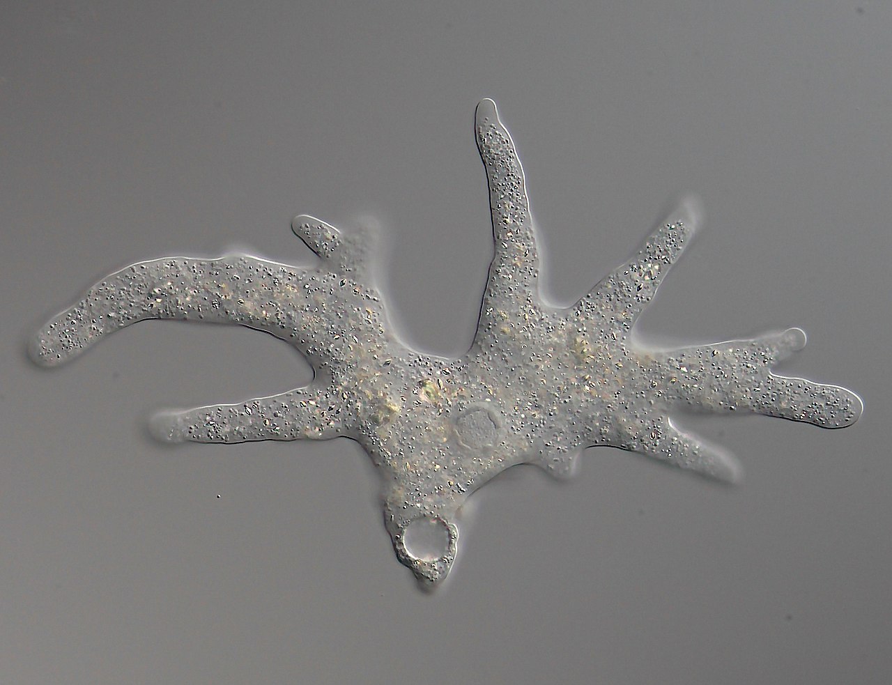

Structure and mechanism
At the core of a pseudopodium is a flow of cytoplasm and a reorganized cytoskeleton. Actin filaments polymerize near the membrane, producing protrusive force that extends the projection; myosin-driven contraction elsewhere in the cell can pull the cell body forward. The cytoplasmic flow and supporting filament network behave much like a deformable mesh: imagine a net being filled and stretched by a soft material or gelatin, allowing the shape to change rapidly. Chemical and mechanical signals at the leading edge guide where protrusions form and how they stabilize.
Types of pseudopodia
Pseudopods vary in shape and persistence. These morphological distinctions are useful for identifying organisms and understanding their behaviour:
- Lobopodia: Broad, rounded bulges common in classic amoebae used both for locomotion and engulfing prey.
- Filopodia: Thin, needle-like spikes that probe the environment and can act as sensory or exploratory structures.
- Reticulopodia: Networks of interconnecting pseudopods that form irregular nets; typical of some foraminiferans and used for trapping particles.
- Axopodia: Very slender, supported by axially arranged microtubules; they can rapidly retract and are seen in groups such as radiolaria and heliozoans.
Functions and examples
Beyond locomotion, pseudopodia are central to phagocytosis — the process by which cells surround and internalize food, debris or microbes. Many protists capture prey by flowing cytoplasm around items and sealing them in internal vacuoles. In multicellular animals, cells of the immune system such as the white blood cell use pseudopod-like extensions to migrate through tissues and engulf pathogens. Pseudopod dynamics also contribute to wound healing, development, and tissue surveillance in higher organisms.
History, terminology and important distinctions
Observations of crawling cells and shape-changing microorganisms date back to early light microscopy; the term pseudopodium reflects its appearance as a "false foot" (from Greek roots meaning "false" and "foot"). Modern microscopy and cell biology have revealed the molecular players—actin, associated regulatory proteins and membrane trafficking—that drive protrusion formation. While related structures such as lamellipodia and invadopodia are specialized, the broad term pseudopodium emphasizes a temporary, cytoplasm-driven projection rather than a permanent organelle.
Notable facts and context
Pseudopodial behavior spans a spectrum: some cells form short, blunt lobes for steady crawling, others extend long filaments to sense gradients. Their study links ecology (how single-celled organisms find food), immunology (how phagocytes respond to infection) and cell biology (how the cytoskeleton generates force). For further introductory resources see general references on cell locomotion and protist biology (cytoplasm overview, locomotion modes, membrane dynamics). Additional reading can be found through categorized entries on eukaryotic cell structure, cell types, and historical summaries of microscopic discoveries (Amoeba examples, network analogies, gel-like cytoplasm, flagella comparison, cilia comparison, immune cell movement, axopodial organisms).
Related articles
Author
AlegsaOnline.com Pseudopodia: cytoplasmic extensions for movement and feeding Leandro Alegsa
URL: https://en.alegsaonline.com/art/79821