Cilia: structure, function, and biological importance
Cilia are hairlike projections on eukaryotic cells that enable movement, fluid flow, and sensing. This article explains their structure, types, roles, history, and clinical relevance.
Cilia (singular: cilium) are slender, microscopic projections that extend from the surface of many eukaryotic cells. Often described as hairlike organelles, they are anchored by a basal body and built around a core of microtubules. Cilia occur singly or in large numbers and are broadly categorized by function into motile cilia, which generate movement or move fluid, and non-motile primary cilia, which act primarily as sensory and signalling hubs. For a general definition see organelle and for cellular context see eukaryotic.
Image gallery
4 Images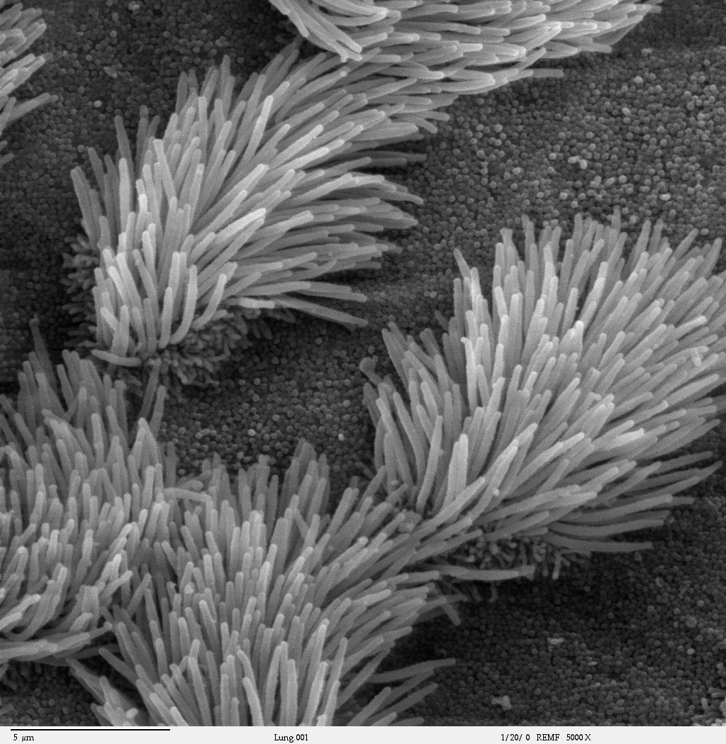


Structure and principal parts
The internal scaffold of a typical cilium is the axoneme, a bundle of microtubules arranged in conserved patterns. Motile cilia usually display a "9+2" arrangement: nine outer doublet microtubules surrounding a central pair. Primary (non-motile) cilia often have a "9+0" arrangement lacking the central pair. Dynein motor proteins attached to the outer doublets produce force by sliding microtubules against one another, producing bending. The basal body, derived from a centriole, anchors the cilium to the cell and organizes its assembly. Many molecular machines, including intraflagellar transport (IFT) complexes, shuttle building blocks along the axoneme during construction and maintenance.
Types and examples
- Motile cilia: beat rhythmically to move cells or move fluid across tissues. They are abundant on protist protists and on cells of multicellular animals. Classic single-celled examples are the ciliates and species such as Paramecium, which use hundreds of cilia for swimming.
- Primary (non-motile) cilia: usually one per cell, serve as antennae for biochemical and mechanical signals. These cilia are central to signalling pathways and to sensing the extracellular environment.
Functions and biological importance
Motile cilia clear mucus and debris from respiratory airways, move eggs through the oviduct, and help establish fluid flow in developing organs. For example, epithelial cilia in the digestive system and the trachea of the lungs coordinate fluid transport and protection. Primary cilia detect chemical and mechanical cues and participate in key developmental signalling pathways, such as Hedgehog signalling. Because they influence cell polarity, proliferation and tissue patterning, cilia are important in embryonic development and adult physiology.
History and scientific development
Observations of ciliary motion date back to early light microscopy, when naturalists first saw beating appendages on microscopic organisms. In the 20th century, electron microscopy revealed the conserved microtubule architecture that distinguishes different cilia types. Subsequent molecular and genetic work identified dynein motors, intraflagellar transport proteins and many regulatory factors that guide assembly and function.
Clinical relevance and distinctions
Defects in cilia form or function cause a range of human disorders collectively called ciliopathies. Examples include primary ciliary dyskinesia, which impairs respiratory clearance and fertility due to defective motile cilia, and syndromic conditions that affect the kidney, brain, eye or skeleton when primary cilia signalling is disrupted. It is important to distinguish eukaryotic cilia and flagella—similar in structure and evolutionarily related—from bacterial flagella, which are mechanically and compositionally different. Research into cilia continues to connect basic cell biology with developmental biology and medicine.
Notable facts: cilia and flagella in eukaryotes are sometimes grouped under the term undulipodia; intraflagellar transport is essential for their assembly; and both single-celled organisms and multicellular tissues exploit cilia for diverse tasks from locomotion to sensory perception.


Secondary cilia
Structure and occurrence
Each cilia is surrounded on the outside by a plasma membrane. Inside the cytoplasm, as a special formation of the cytoskeleton, lies the so-called axoneme consisting of bundles of fine hollow tubulin fibers, the microtubules. In secondary cilia, these are arranged according to the 9×2+2 pattern: In the middle of the cilium, two single central tubules (singletons) lie in a sheath-like envelope (central sheath). These two central singlet tubules are surrounded in a circle by nine double tubules (doublets), each consisting of an A tubule and a B tubule fused to it.
At each A-tubule there are pairs of arm-like structures (dynein arms), which are directed towards the B-tubule of the neighbouring doublet. The dublets arranged in a circle are connected to each other in a ring by nexin binding members as well as to the enveloped central tubules by so-called radial spokes.
All twenty microtubules originate from a basal corpuscle, the formation center of the cilia at the base of the cilia. Fibers of the transition zone anchor this kinetosome in the cell membrane and separate the membrane-enveloped cilia with the axoneme from the rest of the cell body in such a way that one can speak of compartmentalization.
The beat of a secondary cilia as an active movement is produced by energy-requiring tensions of the microtubules inside the cilia. The movement process can be described as a sliding filament mechanism. Arms of dynein, always anchored to the A-tubule, make contact with the B-tubule of the next neighbouring tubule with their tips and can cause an ATP-dependent displacement of the doublets towards each other, so that a bending is produced. Nexin, a highly extensible protein, holds adjacent doublets together during this sliding process.
Such kinocilia are often formed in large numbers on the cell surface of unicellular or multicellular organisms. Flagella and cilia are also scientifically summarized under the generic term undulipodia (singular undulipodium), due to the same construction principle.
Some ciliates possess groups of cilia that are interconnected. Such groupings of projections are also called cirrus (Latin for curl).
Occasionally, cilia are not distinguished clearly enough from microvilli. These, however, do not carry a scaffold of microtubules, but are equipped with actin filaments, furthermore of a different origin and usually only passively mobile. Microvilli, not cilia, are found for example in the intestines of mammals, where they serve to increase the surface area. Here, the food pulp is moved by the peristalsis of muscle cells. Another example of cell processes that are wrongly called cilia are the formations of hair cells in the inner ear. The stimulus-receiving processes of these sensory cells, formerly called stereocilia, are also microvilli and are therefore now called stereovilli. A cilium exists in the human hair cell only in the embryonic anlage and degenerates during development.
The cilia should not be confused with the flagella of bacteria. These are much slimmer, consist entirely of protein (flagellin) and are not surrounded by a membrane. Their mode of operation is also based on a completely different principle (namely a rotation similar to a ship's propeller).
Movement and function
Kinoceils are rarely found alone, but usually in larger numbers and often in rows or fields on a cell. The coordinated, oar-like beating of the motile cilia serves the following functions:
- Locomotion of the individual cell, as in ciliates, numerous larval stages of smaller, water-dwelling animals or also the sperm of higher animals.
- swirling of particles of food
- Transport of particles and fluids within an organism, for example through the ciliated epithelium in the airways a transport of mucus and foreign substances from the bronchi or through ciliary epithelial cells in the fallopian tube the transport of an ovum.
Cinematic cilia are, so to speak, flexible miniature rudders which, in contrast to flagella, beat uniplanarly (in one plane). The plane and direction of stroke are fixed for each cilia. During the powerful stroke the cilium is almost stretched. The slower recoil is curved, with a bending wave running from the base of the cilia to the tip of the cilia, returning the cilium to its original position when there is little resistance from the surrounding medium. A curve in space can be traversed at the same time.
Each cilia of a cilia row beats a fraction later than the previous one. This is called a metachronous movement. The collective movement is wave-like, comparable to a field of grain swaying in the wind.
The beat frequency of a cilium can be between 5 and 20 Hz, depending on environmental conditions. There are factors that can accelerate the frequency, such as heat or some medications. Other factors, however, inhibit the frequency or even cause it to stop, such as nicotine or a bacterial infection.


Primary cilia
In contrast to the mostly actively movable, "motile" secondary cilia, primary cilia are mostly only passively movable. As a rule, only one such "non-motile" cilia exists per cell, which is formed according to the 9×2+0 scheme - the central pair is thus missing.
Almost all vertebrate cells possess a single nonmotile cilium, also called the "primary cilium", which has long been neglected in research. These primary cilia often represent a sensitive extension of the cell. Specialized structures have also evolved from these nonmotile cilia; for example, the outer segment of photoreceptor cells in the eye is connected to the inner segment by a specialized cilium called the connecting cilium.
In contrast, olfactory cells, as sensory cells of smell in vertebrates at the same time olfactory nerve cells, each have up to twenty nonmotile cilia with special olfactory receptors; however, these special nerve cell processes are structured according to the 9×2+2 scheme of secondary cilia.

Questions and answers
Q: What is a cilium?
A: A cilium is an organelle found in eukaryotic cells.
Q: What are the two types of cilia?
A: The two types of cilia are motile cilia and non-motile, or primary cilia.
Q: What is the function of motile cilia?
A: Motile cilia beat against fluid outside the cell and are found on protist ciliates like Paramecium. They are how Paramecium moves around. They are also found on the epithelial cells of many internal organs of metazoans, such as the digestive system and the trachea of the lungs.
Q: What is the function of non-motile, or primary cilia?
A: Non-motile, or primary cilia, typically serve as sensory organelles.
Q: What is undulipodia?
A: Undulipodia is a group of organelles that includes cilia and flagella.
Q: Are eukaryotic cilia and flagella structurally identical?
A: Yes, eukaryotic cilia are structurally identical to eukaryotic flagella, although distinctions are sometimes made according to function and/or length.
Q: Where are motile cilia found?
A: Motile cilia are found on protist ciliates like Paramecium, as well as on the epithelial cells of many internal organs of metazoans, such as the digestive system and the trachea of the lungs.
Related articles
Author
AlegsaOnline.com Cilia: structure, function, and biological importance Leandro Alegsa
URL: https://en.alegsaonline.com/art/20372
Sources
- hhmi.org : "The importance of being cilia"
- encyclopedia.com : A Dictionary of Biology
- ncbi.nlm.nih.gov : "Cilia, flagella, and microtubules"
- doi.org : 10.1083/jcb.91.3.125s
- pubmed.ncbi.nlm.nih.gov : 6459327