Flagellum — cellular locomotor and sensory appendage
A flagellum is a long, whip-like appendage used by many cells and microorganisms for propulsion and sensing. Prokaryotic and eukaryotic flagella are structurally distinct and serve varied biological roles.
A flagellum (plural: flagella) is a slender, often whip-like projection that extends from the surface of many cells and single-celled organisms to produce movement or environmental sensing. In eukaryotic cells the core of a flagellum contains an ordered bundle of microtubules and is typically covered by the cell's plasma membrane; motion is generated by sliding between these microtubules. Flagella propel organisms through fluids by coordinated bending waves, producing a characteristic whip or whip-like motion, and are essential for the locomotion of many protists, sperm cells and some algae. They also play roles in surface attachment and environmental sensing in a wide range of organisms.
Image gallery
9 Images


Structure and mechanics
Eukaryotic flagella are built around an axoneme, a cylindrical array usually organized in a 9+2 pattern of microtubule doublets and a central pair; movement arises from motor proteins that cause sliding between these microtubules. The base of the flagellum is anchored in a basal body that resembles a centriole and organizes assembly. In contrast, bacterial flagella are molecular rotary devices made from different protein subunits and driven by ion gradients; although both types are called "flagella," they are not homologous and work by fundamentally different mechanical principles.
Types, distinctions and terminology
Biologists distinguish eukaryotic flagella from smaller, often more numerous cilia mainly by length and beating pattern: flagella are usually longer and fewer per cell, while cilia are short and occur in arrays. Because of their close structural similarity some researchers have proposed grouping protists that bear either structure under a common category such as protists with undulating appendages; historical classifications have varied, including proposals for a separate Phylum or group for certain ciliated organisms like the Ciliates.
Origin, evolution and cellular context
Flagella are examples of cellular organelles or appendages that work together with other intracellular systems. In eukaryotes flagella operate alongside other organelles such as mitochondria and plastids, which themselves have well-supported evolutionary origins from once-independent prokaryotes such as bacteria and archaea. The evolutionary history of flagella is complex: eukaryotic flagella evolved within the eukaryotic lineage and are structurally distinct from bacterial flagella, so their deep origins remain an active field of study.
Functions and biological importance
- Locomotion: flagella enable swimming or gliding of single-celled organisms, sperm, and some multicellular cells.
- Sensory roles: flagella can detect chemical and physical cues and help cells respond to their environment.
- Ecological and medical relevance: bacterial flagella contribute to motility and colonization, affecting nutrient acquisition and pathogenicity.
Because the word "flagellum" covers mechanically diverse structures, it is useful to specify whether one is discussing eukaryotic, bacterial or archaeal appendages. For general background on cellular organelles and related classifications see resources on cell structure and organelles. For further reading and visual guides follow introductory materials linked in online biology collections and specialized reviews (eukaryotes, cell membrane).
Notable facts: eukaryotic flagella are typically surrounded by the cell membrane and exhibit bending patterns such as an S-shape; bacterial flagella rotate like propellers. Understanding these differences is important in cell biology, microbiology and biomedical research.

The flagella of prokaryotes
Structure
Bacterial flagella are extracellular, helical filaments ("filaments") that are anchored to the cell membrane(s) and cell wall via a "hook" with a motor complex. The flagella, including the hook and motor complex, are composed entirely of proteins. The diameter of the filaments in most flagella is about 15-20 nm, and they are hollow. Because of their small diameter, they can only be made visible by dark-field microscopy and electron microscopy, and not by normal light microscopy, but there are special staining procedures that thicken them enough to make them visible by light microscopy.
During the assembly of filaments, the protein molecules (flagellin) are transported through the hollow channel of the flagella to the outer end where they are attached. If there is a sufficiently large supply of flagellin in the cell, the assembly of a filament can happen very quickly.
The flagella of archaea are functionally similar to those of bacteria, but consist of different proteins and a different motor complex that is driven by ATP.
Flagellation types
According to arrangement and number of flagella, different types of flagella are distinguished (in the order of descending swimming speed):
- holotrich: numerous flagella are evenly distributed over the entire cell surface and cover the entire body surface
- peritrichous: many flagella are scattered evenly over the cell surface.
- polytrich-bipolar: the flagella are in two opposite groups at the cell poles. (also called amphitrichous).
- polytrich-monopolar: the flagella are in a group at one of the cell poles (also called lophotrich).
- monotrich: The cell has only one flagellum.
- polar: The flagellum or flagella are located at one or both poles of the cell.
- lateral, lateral flagella: flagella are located laterally, not at the poles of the cell.
Lateral flagellation is often not associated with high swimming speeds, but it offers the advantage that the bacterium can more easily squeeze into obstacles such as highly viscous liquids or gaps between solids.
Method of movement
Due to their helix, the flagella act similar to a propeller. The motor complex converts a difference in concentration of protons between the two sides of the inner cell membrane into a rotary motion of the coiled filament sitting on a curved "hook" and thus follows a similar construction principle as ATP synthase. The flagellar mechanism represents the only truly rotating joint known in all of biology to date. The rotation frequency is around 40-50 Hz.
The direction of the flagellar rotation caused by the motor in combination with the winding direction of the flagellar coil determines whether a push or a pull is exerted on the bacterial body. The direction of the rotation caused by the motor can be reversed in a very short time, so that push and pull can change quickly.
As a rule, the direction of rotation of the flagella is such that they push. This means that they are at the posterior end in monopolar flagellated bacteria. The bacterial body thereby rotates (more slowly) in the opposite direction (conservation of angular momentum).
In bipolar flagellated bacteria, the flagella of the two ends rotate in opposite directions. Thus, the flagella of the posterior end have a pushing effect, the flagella of the anterior end are bent backwards and rotate around the anterior end of the bacterial body, thus increasing the thrust. If the direction of rotation of the flagella is reversed, the filaments fold over, the rear end of the bacterium becomes the front end and the front end becomes the rear end, the bacterium swims in the opposite direction.
The flagella of peritrichous flagellated bacteria rotate in the same direction and usually in such a way that they push. In doing so, they unite to form a backward coiled bundle, also known as a "flagellar pigtail", which pushes the bacterium forward. If the direction of rotation of the flagella of peritrichous flagellated bacteria is reversed, the individual flagella straighten radially away from the bacterial body and their pulling action on the bacterial body cancels out on average, causing the bacterium to tumble in random motion in one place.
Reversal of the direction of flagellar rotation and the associated change in the direction of movement plays an important role in taxa (see for example chemotaxis).


The scourges of the eukaryotes
Structure
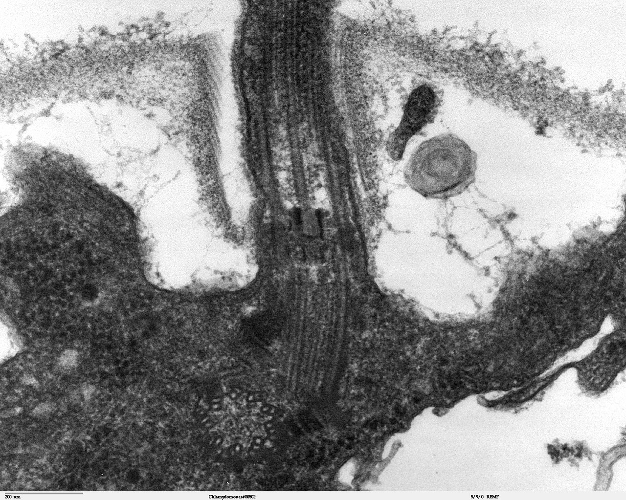
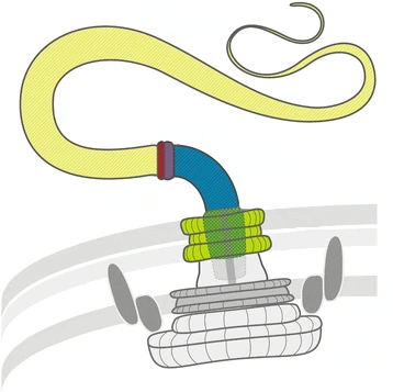
The flagella of eukaryotes are filamentous structures that project outward from the body into the surrounding medium and are surrounded by the cell membrane and filled with cytoplasm. Inside them lie microtubules in a special arrangement called 9×2+2; Nine double microtubules form a circle in cross-section, with two single microtubules in the middle. The double microtubules each consist of one complete microtubule (A-tubule) and one incomplete one (B-tubule). On the A-tubule, pairs of protein arms called dynein arms are located at equal height intervals, approximately every 20 nm. This microtubule arrangement is called the 9×2+2 structure, and the entire microtubule bundle is called the axoneme. This structure is stabilized by various bridging proteins (mainly nexin). At the base of the flagellum, where it merges into the cell body, there is a basal apparatus called the blepharoplast or kinetosome, which structurally resembles a centriol. It consists of nine triple microtubules in a circle (9×3 structure) that is transverse to a second 9×3 structure of the same structure. It is often referred to as a centriol. At the free end, the eukaryote flagella are pointed. Their diameter is about 250-300 nm, their length a few micrometers to more than 150 µm.
An illustrative example of flagellum-bearing cells is provided by spermatozoa. The movement goes in a wave with constant amplitude from the base to the tip of the flagellum.
Together with the cilia, the flagella of eukaryotes are also called undulipodia.
Method of movement
According to the current state of knowledge, the shape change required for hydrodynamic effectiveness is achieved by sliding of the double fibrils in opposite directions. The energy for this is to be provided by the dynein arms, namely by hydrolytic cleavage of phosphate from ATP. The protein dynein, which forms the dynein arms, has ATPase activity. Microtubule sliding results in a change in the shape of the flagellum.
The shape changes of the flagella vary depending on the type of flagella. They may consist in a wave (undulation) running over the flagellum in a plane or in the form of a helix with circular to elliptical movements; they may also consist in a flagellar stroke, whereby the flagellum curves in one direction and, as it were, infiltrates the medium and strikes outstretched in the opposite direction, thus exerting a force. Flagella which move in the manner last described are called cilia. They are usually shorter than other flagella, and are arranged in greater density on the surface of cells and tissues.
The consequence of the flagellar movement can be a locomotion of the individual, but it can also result in a movement of the adjacent medium or nearby particles when the individual is at rest. Examples of locomotion of the individual: freely moving ciliates, flagellates, spermatozoa. Examples of locomotion of the adjacent medium or particles: sessile ciliates, ciliated epithelium in the trachea of animals.
Variations, flagellation types
In some unicellular algae and protozoa, the flagella are occupied by many lateral short filaments called mastigonemes or cilia and are called ciliated flagella. The mastigonemes may occur in one row (stichonematic) or in two rows (pantonematic).
If a cell carries several flagella, it is called an isocont flagellum if they are of the same kind, as for example in green algae Cells with different flagella are called heterocont or anisocont, where usually a long, forward directed ciliated flagellum serves as a traction flagellum and a short smooth trailing flagellum is directed backwards, as for example in the heterocontae. Cells without flagella are called acont in distinction to the flagellated forms.
According to the place of insertion of the flagellum, a distinction is made between acrokont (at the front end, traction flagellum), pleurokont (lateral) and opisthokont (at the rear end, thrust flagellum).


Questions and answers
Q: What is a flagellum?
A: A flagellum is a long, whip-like structure that helps some single celled organisms move.
Q: What is the composition of a flagellum?
A: A flagellum is composed of microtubules.
Q: How does a flagellum help propel cells and organisms?
A: A flagellum helps propel cells and organisms in a whip-like motion.
Q: What is the motion of the flagellum of eukaryotes?
A: The flagellum of eukaryotes usually moves with an “S” motion and is surrounded by cell membrane.
Q: What are cilia?
A: Cilia are cell organelles that are structurally almost identical with flagella.
Q: What is Protista?
A: Protista is a collection of disparate single-celled forms and a useful term for now.
Q: What is the origin of cell organelles such as cilia and mitochondria in eukaryotes?
A: All or most of these organelles have their origin in once-independent prokaryotes (bacteria or archaea), and the eukaryote cell is a 'community of micro-organisms' working together in 'a marriage of convenience'.
Related articles
Author
AlegsaOnline.com Flagellum — cellular locomotor and sensory appendage Leandro Alegsa
URL: https://en.alegsaonline.com/art/34971
Sources
- doi.org : 10.1038/249073a0
- pubmed.ncbi.nlm.nih.gov : 4598030
- doi.org : 10.1038/325637a0
- doi.org : 10.1038/245380a0
- pubmed.ncbi.nlm.nih.gov : 4593496
- doi.org : 10.1146/annurev.mi.19.100165.000321
- pubmed.ncbi.nlm.nih.gov : 5318439
- doi.org : 10.1146/annurev.micro.57.030502.091014
- pubmed.ncbi.nlm.nih.gov : 14527279
- doi.org : 10.1159/000094053
- pubmed.ncbi.nlm.nih.gov : 16983194
- doi.org : 10.1007/s10541-005-0065-8
- pubmed.ncbi.nlm.nih.gov : 15627373
- encyclopedia.com : A Dictionary of Biology