
Myoglobin: structure, function, physiology and clinical significance
Myoglobin is a heme-containing, oxygen-binding protein in muscle that stores and facilitates oxygen diffusion; it has roles in physiology, meat color, diving adaptations and as a clinical marker of muscle injury.
Overview
Myoglobin is a small, heme-containing protein found primarily in the muscle cells of vertebrates. It reversibly binds oxygen, serving as an intracellular oxygen reservoir and helping to shuttle oxygen from the bloodstream to mitochondria during periods of high demand. Because of its pigmenting heme group it also contributes to the red or dark color of muscle tissues such as beef and lamb.
Image gallery
10 Images




Structure and biochemical properties
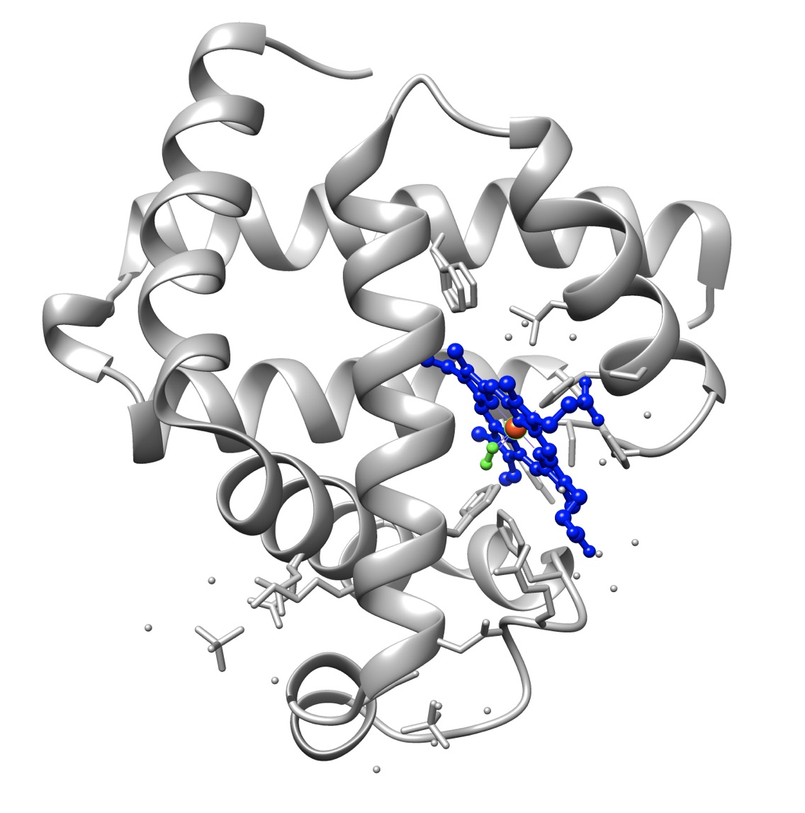
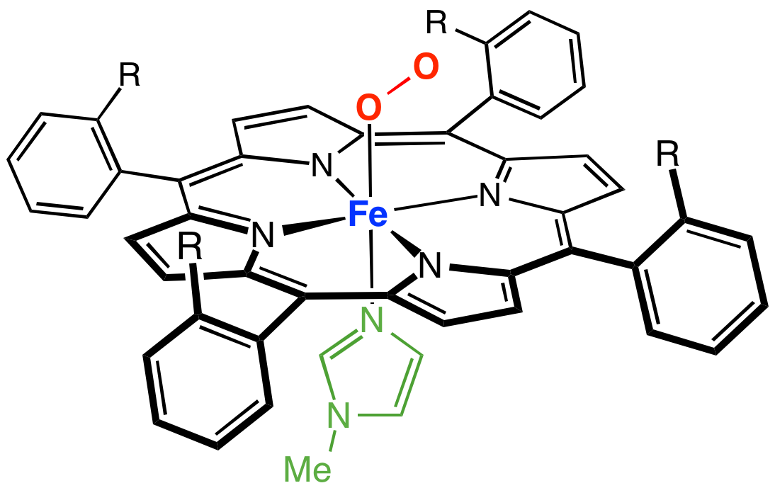
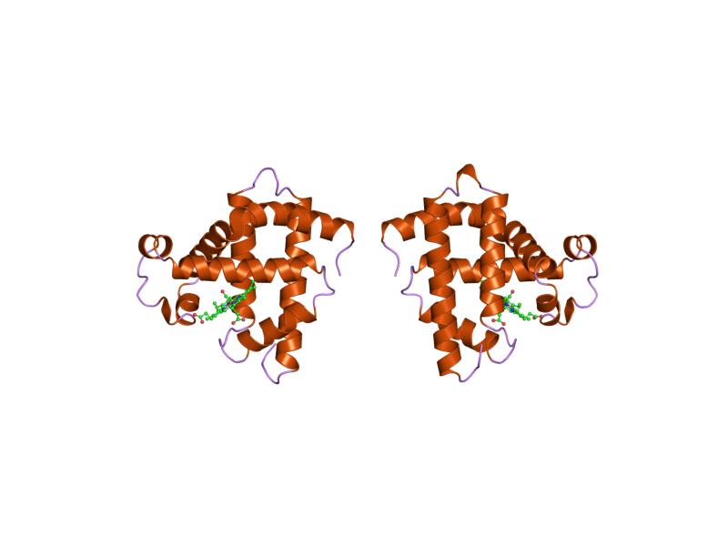
Myoglobin belongs to the globin family and displays the characteristic globin fold of eight alpha helices arranged around a hydrophobic pocket that holds a prosthetic heme (a planar porphyrin ring coordinated to a central iron atom). Human myoglobin is a single polypeptide chain of 153 amino acids and has an approximate molecular mass of 17 kDa. In the ferrous state (Fe2+) it binds oxygen to form oxymyoglobin; oxidation to ferric iron (Fe3+) produces metmyoglobin, which cannot carry oxygen and is associated with the brown color of aged meat.
Physiological role and distribution
Myoglobin is concentrated in oxidative (slow-twitch) muscle fibers that rely on sustained aerobic metabolism. It both stores oxygen for short-term use and facilitates intracellular diffusion of oxygen to mitochondria when blood flow is limited. Levels of myoglobin vary between species and lifestyles: many mammals possess it, and diving mammals such as seals and other marine mammals typically have particularly high myoglobin concentrations to support prolonged dives. These elevated levels increase the total oxygen reserve available to working muscles.
Clinical significance and laboratory testing
When muscle is damaged, myoglobin can be released into the blood and detected in the serum. A rise in serum myoglobin may indicate acute muscle injury, including skeletal muscle trauma, epileptic seizures, or cardiac muscle injury such as myocardial infarction. Because myoglobin appears in blood early after injury but clears rapidly, it has been used as an early marker for muscle damage; however, it is less specific for heart damage than cardiac troponins. Very high concentrations of circulating myoglobin can precipitate in kidney tubules and contribute to acute kidney failure in rhabdomyolysis, making prompt recognition and treatment important.
Applications, research and notable facts
- Model protein: Myoglobin was the first protein whose three-dimensional structure was solved by X-ray crystallography, a landmark achievement in structural biology led by researchers such as John Kendrew; it remains a classic model for studies of protein folding, ligand binding and spectroscopy.
- Meat industry: Myoglobin content largely determines the redness of meat—higher myoglobin yields darker red muscle, lower myoglobin yields pale or white meat.
- Toxicology: Carbon monoxide binds to the heme iron with high affinity, forming carboxymyoglobin and impairing oxygen transport in muscle.
- Laboratory use: Myoglobin is measured by immunoassays and spectrophotometric methods; it is also used experimentally to study oxygen binding kinetics because its oxygen-binding curve is non-cooperative and hyperbolic, unlike the sigmoidal curve of hemoglobin.
Distinctions and historical notes
Although functionally related to hemoglobin, myoglobin is a monomeric oxygen storage and diffusion protein rather than a tetrameric oxygen transporter. Its single-chain architecture and well-characterized heme environment made it ideal for early protein chemistry and structural studies. Variants and rare myoglobin-related disorders are uncommon; most clinical attention focuses on its role as a marker of muscle injury and on the renal complications that may follow massive myoglobin release.
For further reading and resources, see specialized entries on the chemistry of the heme group and on diagnostic markers in cardiology and nephrology: protein chemistry, oxygen transport, muscle pigments, porphyrins, iron biochemistry, muscle physiology, mammalian adaptations, seal physiology, marine mammal biology, serum markers, blood testing, myocardial infarction, seizure-related injury, acute kidney injury.
Related articles
Author
AlegsaOnline.com Myoglobin: structure, function, physiology and clinical significance Leandro Alegsa
URL: https://en.alegsaonline.com/art/67983
Sources
- doi.org : 10.1242/jeb.01172
- pubmed.ncbi.nlm.nih.gov : 15339940