Myosin: the actin-based motor proteins of eukaryotic cells
Myosins are a large family of ATP-dependent motor proteins that bind actin to produce force and motion in eukaryotic cells, with conserved structure and many specialized classes.
Myosin denotes a broad family of motor proteins that convert chemical energy into mechanical work by coupling ATP hydrolysis to movement along actin filaments. Found throughout eukaryotic life, myosins drive processes ranging from muscle contraction to intracellular transport and cell motility. They are studied both as fundamental components of cell biology and as targets in medical and biotechnological research. For a general overview see protein family descriptions and cellular context in eukaryotic cell references.
Image gallery
10 Images


Structure and mechanism
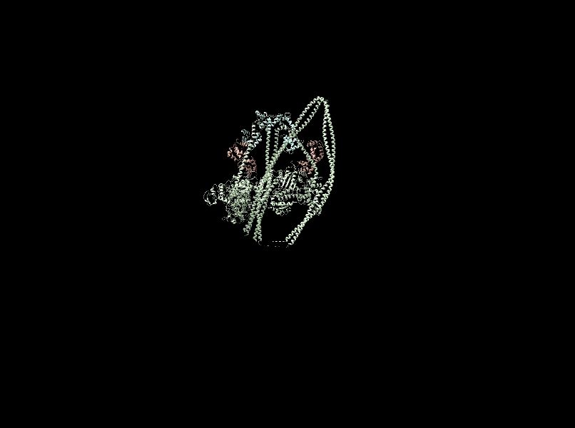
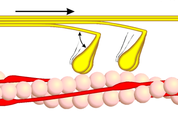
Myosin molecules share a conserved modular design: a globular head (motor) domain that binds actin and nucleotides, a neck or lever arm that amplifies small conformational changes, and a variable tail that mediates dimerization or cargo binding. The head contains the ATP-binding pocket and the actin interface; hydrolysis of adenosine triphosphate (ATP) and subsequent product release power a conformational "power stroke" that produces movement. This basic chemo-mechanical cycle underlies myosin function in diverse cellular tasks.
Types, diversity and genes
Multiple classes of myosin (conventionally numbered I, II, V, VI, etc.) perform distinct roles. Conventional myosin II forms bipolar filaments that slide actin in muscle and contractile networks, while unconventional myosins (e.g., myosin I, V, VI) act as monomers or dimers to carry vesicles, organize membranes, or participate in endocytosis. A large repertoire of myosin genes exists across species; comparative genomics and expression studies catalog these variants and their tissue-specific roles—see genetic and annotation resources via myosin gene lists and functional summaries at structure and function resources.
Actin–myosin interactions are highly conserved: for instance, skeletal myosin II from a mammal can interact productively with actin from distantly related eukaryotes, underscoring a conserved interface. Experimental assays—biochemical ATPase measurements, in vitro motility assays, and single-molecule experiments—have elucidated force production, step size, and duty cycle across classes. For additional molecular context see actin-associated studies.
Biologically, myosins power macroscopic contraction in muscle, generate cortical tension during cytokinesis, transport organelles and mRNA, and remodel the cytoskeleton during cell migration. Mutations in particular myosin genes are implicated in human diseases such as cardiomyopathies, hearing loss, and some forms of kidney disease, so myosins are medically significant as well as mechanistically interesting.
Historically, myosin was recognized through muscle physiology and later shown to be an ATPase that binds actin; advances in structural biology, single-molecule biophysics, and genetics have since expanded understanding of its diversity and regulation. Ongoing research explores regulatory light chains, post-translational control, and engineered myosins for nanotechnology applications.
Notable distinctions include the varied duty ratios (time spent attached to actin), filament-forming ability, and cargo specificity among classes—properties that tune each myosin to its cellular role. Practical resources and reviews provide deeper technical detail and experimental protocols for researchers and students.
Questions and answers
Q: What are myosins?
A: Myosins are groups of specialized proteins used for muscle contraction and motion in eukaryotic cells.
Q: What do myosins need for energy to function?
A: Myosins need adenosine triphosphate (ATP) for energy to perform their functions.
Q: How many different myosin genes have been discovered in eukaryotes?
A: A large number of different myosin genes have been discovered in eukaryotes.
Q: Is the structure and function of myosin conserved across species?
A: Yes, the structure and function of myosin is strongly conserved across species.
Q: What is an example of myosin's conservation across species?
A: An example of myosin's conservation across different species is that rabbit muscle myosin II will bind to actin from an amoeba.
Q: What is the function of myosins?
A: The function of myosins is for muscle contraction and motion in eukaryotic cells.
Q: What do myosins need ATP for?
A: Myosins need ATP for energy to perform their functions of muscle contraction and motion in eukaryotic cells.
Related articles
Author
AlegsaOnline.com Myosin: the actin-based motor proteins of eukaryotic cells Leandro Alegsa
URL: https://en.alegsaonline.com/art/67988
Sources
- doi.org : 10.1002/cm.10014
- pubmed.ncbi.nlm.nih.gov : 11810692