
Oesophagus (esophagus): structure, function, and clinical significance
The oesophagus (or esophagus) is the muscular tube that conveys food from the pharynx to the stomach; this article explains its anatomy, physiology, development and common disorders.
Overview
The oesophagus, also spelled esophagus and commonly called the gullet, is the muscular conduit that links the throat to the stomach. It lies within the chest and mediastinum and provides the passage for swallowed food and liquids from the pharynx into the stomach, which is part of the gastrointestinal system. In adults the length is typically around 25 centimetres (about 10 inches), varying with body size and posture.
Image gallery
10 Images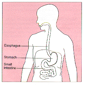




Structure and tissue layers
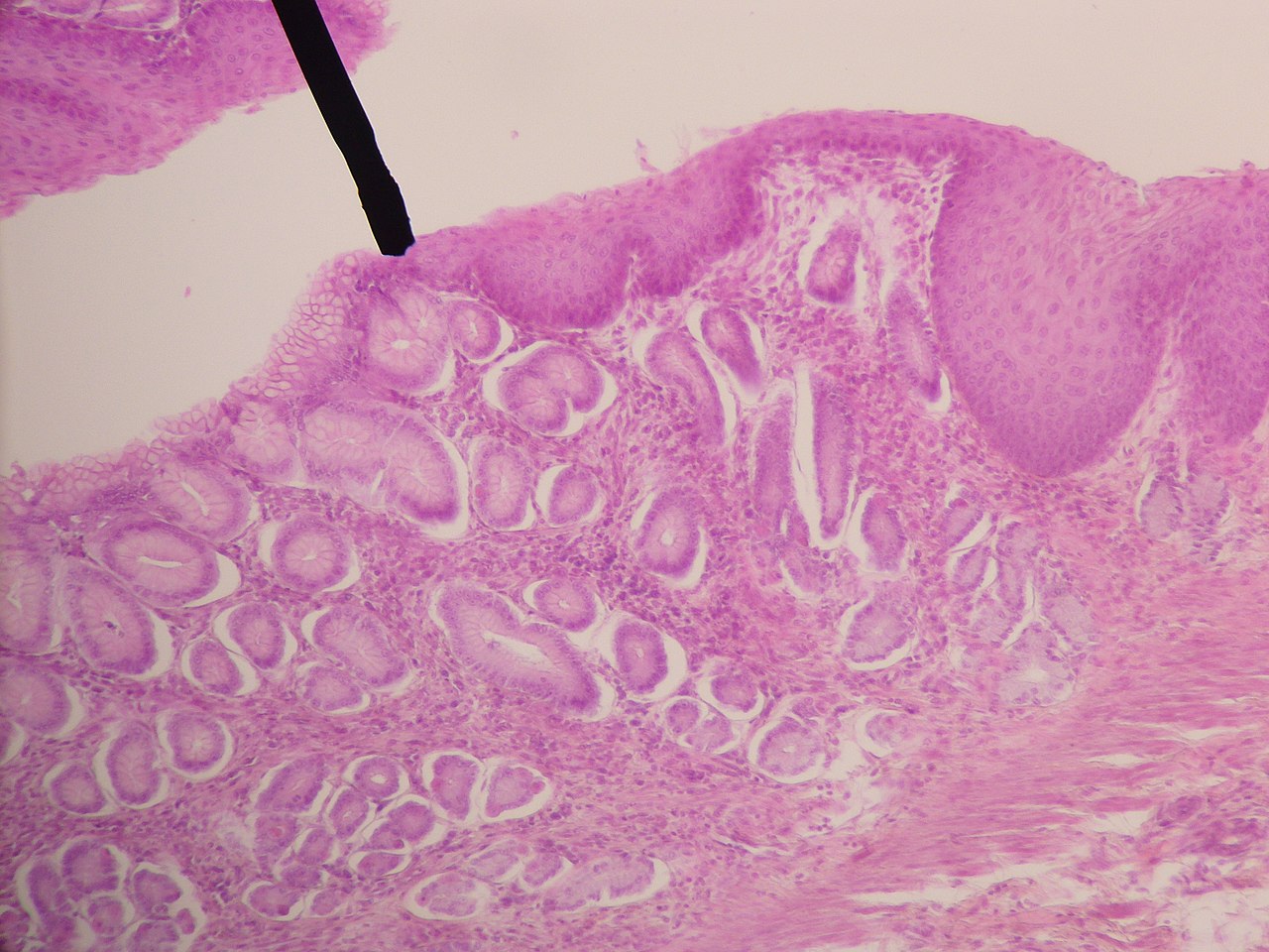
Anatomically the oesophagus is organized into several identifiable regions and layers. From the inside out the wall is composed of mucosa, a supportive submucosa that contains mucus-secreting glands, a muscular layer (muscularis) made up of inner circular and outer longitudinal fibres, and an outer connective tissue layer (adventitia). Proximally the musculature includes striated (skeletal) muscle under voluntary control, while the distal portion is predominantly smooth muscle under autonomic control. Two muscular rings act as sphincters: the upper oesophageal sphincter near the throat and the lower oesophageal sphincter at the stomach entrance.
Function and movement
The primary function of the oesophagus is transport. Swallowing initiates a coordinated sequence of events that moves a bolus of food into the oesophagus and then toward the stomach. This propulsion is accomplished by waves of coordinated muscle contraction and relaxation known as peristalsis. The inner glands and mucous layer keep the lumen lubricated, facilitating smooth passage. The muscular wall itself is composed of organized muscle fibres that generate the contractions responsible for transport.
Development and variation
Embryologically the oesophagus arises from the foregut and matures as neural and muscular control of swallowing and sphincter function develop. Across vertebrates the structure can vary: in some animals certain sections are reinforced for storage or regurgitation, while in humans the tube is primarily a transport organ. The absence of a true serosal covering over much of the oesophagus is a notable anatomical distinction compared with many abdominal organs.
Clinical importance and common conditions
Because the oesophagus is the route for swallowed material and lies close to other thoracic structures, it is frequently implicated in clinical problems. Common conditions include:
- Gastro-oesophageal reflux disease (GERD) and oesophagitis
- Stricture formation and peptic damage
- Barrett's oesophagus, a premalignant change from chronic reflux
- Motility disorders such as achalasia and diffuse oesophageal spasm
- Benign and malignant tumours
Diagnostic approaches used to evaluate symptoms include upper endoscopy, radiographic studies (barium swallow), and manometry to measure pressure and peristaltic activity. Treatments range from lifestyle modification and medication to endoscopic procedures and surgery, depending on the disorder.
Notable facts and distinctions
The oesophagus does not digest food but is essential for delivering the bolus to the stomach, where mechanical and chemical digestion convert it into chyme. Its capacity to relax and expand lets it handle solids, liquids and even larger boluses with coordinated motion. Unlike hollow abdominal organs that are fully covered by serosa, most of the oesophagus is attached to surrounding tissues by adventitia, which affects how infections and cancers spread. For further introductory resources see general references to the gastrointestinal tract and clinical summaries available at medical sites and textbooks (throat and swallowing anatomy, muscle structure, mucosal protection, peristalsis mechanics).
Related articles
Author
AlegsaOnline.com Oesophagus (esophagus): structure, function, and clinical significance Leandro Alegsa
URL: https://en.alegsaonline.com/art/72003