
Peristalsis: coordinated muscular waves in digestion and other organs
Peristalsis is the coordinated contraction and relaxation of muscles that propels contents through tubular organs. This article explains its mechanism, anatomy, examples, control and clinical relevance.
Overview
Peristalsis is a rhythmic, wave-like sequence of muscle contractions that moves contents along a tubular organ. It is most often discussed in the context of the digestive tract but also occurs in other hollow structures such as the ureters and reproductive tubes. The process relies on alternating contractions and relaxations of the muscular wall to push a bolus, liquid, or other material in a preferred direction.
Image gallery
3 Images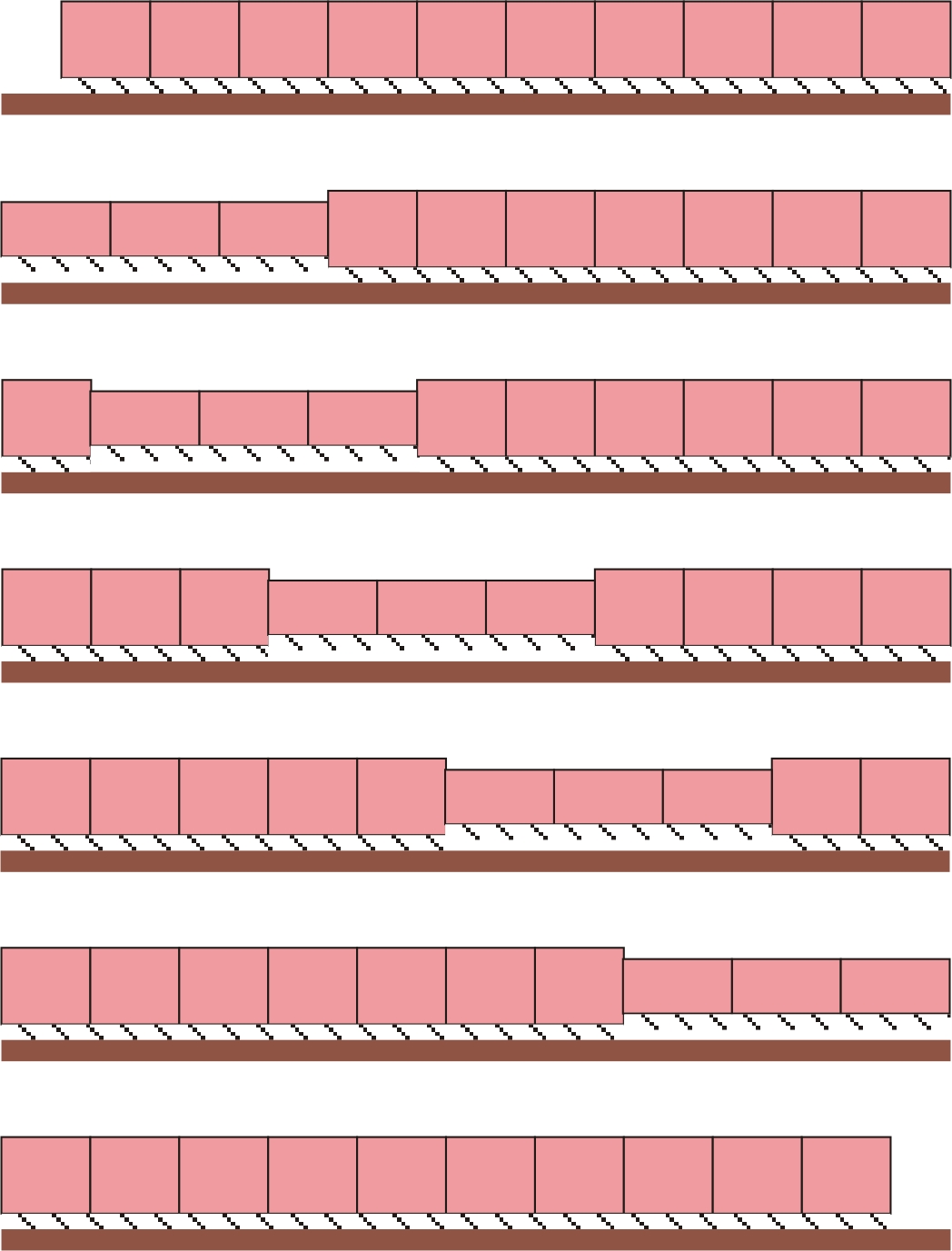
Mechanism and characteristics
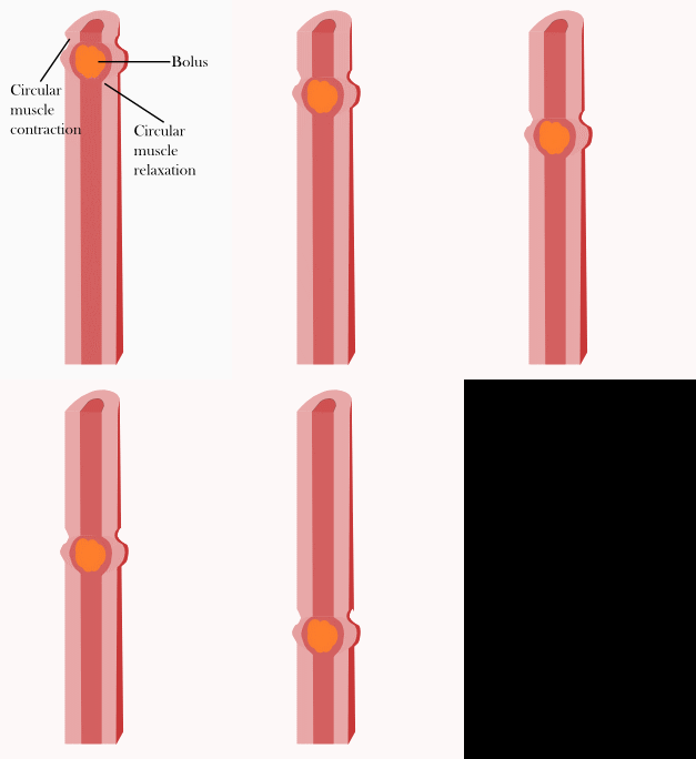
At the tissue level, peristalsis is produced by concentric contraction of circular muscle layers followed by longitudinal muscle changes that shorten and widen the segment. These coordinated actions create a pressure gradient that propels contents forward. The pattern can vary in speed, strength and direction: some segments display strong single waves that clear a lumen, while others produce continuous slower mixing contractions.
Anatomy and muscle types
The muscular work of peristalsis is carried out by specialized muscle layers. In the gastrointestinal tract, an inner circular and outer longitudinal muscle layer act together. In many regions these are smooth muscle, but parts such as the upper esophagus contain skeletal muscle under voluntary control. The term alimentary canal refers to the full digestive tube from mouth to anus; peristaltic activity occurs along much of this pathway, though the pattern and regulation differ by region (alimentary canal).
Neural control and motor patterns
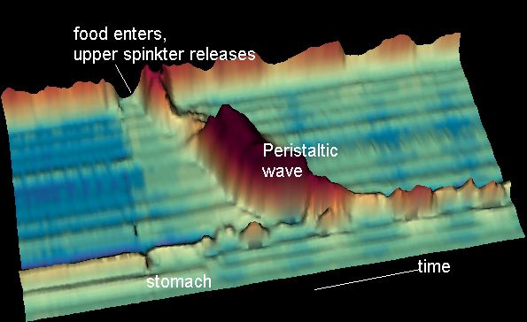
Peristalsis is coordinated primarily by the enteric nervous system, a local network of neurons that can generate reflexes independently of the central nervous system. Autonomic inputs and hormones modulate these reflexes. A distinct motor pattern, the migrating motor complex (MMC), occurs during fasting and travels from the stomach through the small intestine in cyclical sweeps; it is one of several patterns of intestinal motility and differs from the continuous peristalsis seen after a meal. The esophagus provides a familiar example of coordinated propulsion: swallowing triggers an organized peristaltic wave that moves the food bolus downwards (esophagus).
Examples, functions and clinical significance
- Digestive transit: moves swallowed food, mixes luminal contents and advances chyme toward absorption sites.
- Urinary tract: ureteric peristalsis propels urine from kidney to bladder.
- Reproductive tracts: ciliary and muscular movements assist gamete and embryo transport in some species.
Disorders of peristalsis can cause symptoms ranging from difficulty swallowing and reflux to intestinal obstruction and chronic constipation. Conditions such as achalasia involve failure of normal esophageal peristalsis, while postoperative ileus represents a temporary loss of intestinal motility. Diagnostic and investigational tools include manometry to measure pressure waves, transit studies with radiopaque markers, and imaging techniques. Treatments may target underlying causes or use prokinetic medications, dilation, or surgery where appropriate.
Notable facts and distinctions
Peristalsis should be distinguished from segmentation — a mixing movement caused by alternating contractions that do not produce net movement — and from reflex expulsive contractions like vomiting. While the basic mechanical principle is similar across organs, control mechanisms and tissue composition vary: for example, voluntary swallowing engages striated muscle and central pattern generators in the brainstem, whereas much intestinal peristalsis is locally organized by the enteric nervous system and modulated by hormones and autonomic input. For general background and clinical overviews see standard physiology texts and clinical resources (muscle, alimentary canal, esophagus, smooth muscle).
Related articles
Author
AlegsaOnline.com Peristalsis: coordinated muscular waves in digestion and other organs Leandro Alegsa
URL: https://en.alegsaonline.com/art/75844