Stomach
![]()
The title of this article is ambiguous. For other meanings, see Stomach (disambiguation).
The stomach (from Middle High German mage; Ancient Greek γαστῆρ gaster, Latin ventriculus; also Greek Latin stomachus) is a digestive organ of almost all animals. The most primitive form is the gastral cavity of cnidarians, which is also an excretory organ. The stomach is a hollow organ made of muscle tissue, lined internally with a mucous membrane. In contrast to the single-cavity stomach of humans and most animal species, the stomach of ruminants and birds has several demarcated cavity systems (multicavity stomach) as well as highly specialized organs in insects (honey stomach, sucking stomach). Stomachless are e.g. the carp-fish (one interprets this as original adaptation to snail and mussel-food, whose lime-shells made the stomach-acid useless).
The capacity of the human stomach varies from person to person and is about 1.5 litres. In this cavity, the food pulp is mixed with gastric juice, which consists mainly of the protein-splitting enzyme pepsin and hydrochloric acid. The stomach also brings the food pulp to the same temperature, and also stratifies and stores it. When at rest, the glands secrete about 10 ml of gastric juice per hour. When food is ingested, the formation of gastric juice can be increased up to 1000 ml per hour. The formation is controlled both by nerve impulses (especially before food intake) and by hormones. Muscle contractions (peristalsis) transport the food pulp further to the pylorus. This sphincter forms a closure between stomach and intestine and can open about 13 mm wide for the passage of the digested food pulp (chyme). This happens regularly in order to pass the food pulp evenly into the duodenum following the stomach outlet.
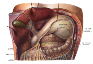
Here the large mesh between the stomach and the transverse colon has been cut and the stomach lifted so that its back and the pancreas behind it can be seen
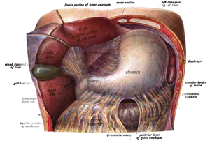
Anatomical drawing of the human stomach and the surrounding structures, the liver has been drawn to the right and upwards
Biological significance
Pre-digestion
In most animals and humans, the actual digestion and absorption takes place in the intestine or, more specifically, in the small intestine. In the stomach, mainly proteins are pre-digested into so-called polypeptide chains. The enzymes necessary for protein digestion, pepsin and cathepsin, have maximum activity due to the low pH value. They are secreted by the cells of the stomach as inactive precursors (pepsinogen) and are only activated by contact with the stomach acid. Fats pass the stomach largely unhindered, but they are softened (liquefied) by peristalsis. Carbohydrates pass the stomach just as unhindered, since the enzymatic cleavage of the α(1-4)-glycoside bond of amylose by α-amylase (from saliva) is inhibited again by the acidic pH in the stomach.
Killing bacteria
The acidic gastric juice effectively prevents the survival of most bacteria and thus protects against infections. For a long time, the doctrine was that in the extremely acidic environment of the human stomach, the growth of microorganisms was in principle not possible. However, with the discovery of Helicobacter pylori in the gastric mucosa in 1983, this view had to be revised. From then on, H. pylori was considered to be the only exception - a bacterium that is able to settle below the protective mucus layer. In 2005/2006, however, scientists at Stanford University were able to prove that the flora (cf. intestinal flora) is considerably more complex than previously assumed: They discovered a total of 128 additional bacterial species colonizing the stomach, including a relative of the extremophilic genus Deinococcus. A total of about ten percent of the identified microorganisms differed significantly genetically from all previously known bacteria.
Incorrectly, the pH value prevailing in the stomach is often compared with concentrated acid. The gastric juice also has a very low pH value of 0.8 to 1.5, but the concentration of hydrochloric acid is lower. However, this low pH value only applies when the stomach is empty. With food intake, the value increases and can rise to 4.5 to 6.5 depending on the buffering capacity of the food pulp.
Regular further digestion
The stomach makes it possible for living beings to get by with a few larger meals per day. It is able to adjust its wall tension to the respective amount of contents (accommodation). The gatekeeper (pylorus) ensures that all substances remain in the stomach for a sufficiently long time and are sufficiently mixed with the digestive juices. The food pulp is then slowly and evenly fed into the intestine.
Structure in humans and animals with monoclavicular stomachs
Anatomy
Seen from the outside, the stomach is divided into the anterior upper surface (paries anterior) and the posterior lower surface (paries posterior), the major curvature (curvatura major), to which the omentum majus is partially attached, and the minor curvature (curvatura minor), to which the omentum minus is attached.
Macroscopically, it is subdivided into the following areas:
- The pars cardiaca (also cardia or cardia, "entrance to the stomach") - with the ostium cardiacum ("mouth of the stomach") - represents the transition between the oesophagus and the stomach.
- The fundus ventriculi (or fundus gastricus, fornix ventriculi, fornix gastricus, "base of the stomach", "dome of the stomach", "vault of the stomach") lies below the diaphragm to the left and cranial of the entrance to the stomach and is filled with air swallowed during food intake. On the X-ray it appears as a gastric bubble.
- The corpus ventriculi (or corpus gastricum, "stomach corpus", "stomach body") makes up the largest portion of the stomach.
- The pars pylorica is narrower than the corpus, the part closer to the duodenum, the canalis pyloricus ("gateway canal"), being even narrower than the more distant part, the antrum pyloricum ("gateway cavity"), also called the vestibulum pyloricum.
- The pylorus ("gatekeeper") is the connection to the duodenum via the ostium pyloricum ("gatekeeper's mouth"). It appears from the outside as a hard constriction and forms the sphincter muscle (Musculus sphincter pylori) towards the inside.
The shape and position of the stomach is not constant, but rather depends on its filling and the position of the person. The pars cardiaca, however, is relatively strongly attached by connective tissue, while the pylorus, which is only attached to the ligamentum hepatoduodenale, lowers when the contents of the stomach are larger, causing the stomach to form the typical hook shape. In women the stomach is usually deeper and steeper than in men.
See also: Blood vessels: Truncus coeliacus and gastric lymph nodes
Histology
Microscopically, the stomach consists of:
- the gastric mucosa (tunica mucosa gastrica), which consists of the laminae epithelialis, propria and muscularis mucosae. It is divided into numerous fields (areae gastricae) by depressions (foveolae gastricae), with a large number of glands (glandulae gastricae).
- a layer of connective tissue (tela submucosa) with blood vessels
- a muscular layer (tunica muscularis gastrica) of smooth muscle consisting of the fibrae obliquae, the stratum circulare and the stratum longitudinale
- a coating of the tunica serosa (peritoneum)
For the secretion of hormones and other secretions in its mucous membrane, the stomach has different types of cells, which are localized in typical places of the stomach, in particular
- in corpus and fundus: accessory cells (form mucus), principal cells (form pepsinogen), parietal or vestibular cells (form hydrochloric acid and intrinsic factor),
- in the antrum: G-cells (produce gastrin).
Embryology
The stomach arises as a spindle-shaped extension of the foregut (the anterior segment of the primitive intestinal tube of the embryo). This is attached to the dorsal and ventral body walls, respectively, by two mesenteries (mesogastrium dorsale and ventrale). The stomach, which initially stands in the longitudinal axis of the embryo, widens on the dorsal side to form the curvatura major and curves on the ventral side to form the flat concave curvatura minor.
The final shape and position of the stomach is formed by different growth of the stomach wall sections. The changes in position are also referred to as "gastric twists" (not to be confused with the disease gastric torsion), although in the process not passive twists but complicated remodelling processes of the stomach anlage take place. The so-called "1st gastric rotation" can be imagined as a twisting around the longitudinal axis to the left by 140 degrees. This causes the large curvature of the stomach to move from the dorsal side to ventrolateral (left ventral), and the small curvature of the stomach to move correspondingly to the right dorsolateral (lateral dorsal). The "2nd gastric rotation" can be rewritten as a rotation around the vertical axis by 90 degrees to the left. This causes the entrance of the stomach to move to the left and the pylorus to move to the right. The "3rd gastric rotation" is again about the longitudinal axis to the right by about 45 degrees. The curvatura major now points to the left and caudoventrally, the curvatura minor to the right and craniodorsally.
gastric blood supply
The stomach is supplied arterially via the coeliac trunk. This short vascular trunk branches off directly from the aorta into three main branches: the common hepatic artery, the sinistral gastric artery and the splenic artery. The stomach is supplied directly by the arteria gastrica sinistra or indirectly by further branches of the other two. The upper part of the small curvature receives its oxygenated blood directly from the Arteria gastrica sinistra. It converges with the gastrica dextra artery, which, originating in the common hepatic artery, supplies the lower part of the small curvature. Both run along the stomach in the lesser reticulum (omentum minus), where they meander along the right side of the stomach. Also originating in the common hepatic artery, the gastroomental artery dextra supplies the lower half of the greater curvature. It converges with the gastroomental sinistra artery, which in turn arises from the splenic artery. Both run at the stomach in the great web (omentum majus). In addition, the splenic artery gives off several gastric arteries, which are responsible for the supply of the fundus, and a posterior gastric artery, which provides blood flow to the posterior wall of the stomach.
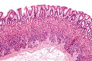
Microscopic preparation of normal gastric mucosa
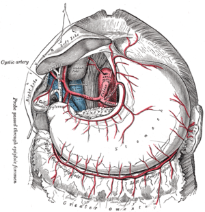
blood supply of the human stomach
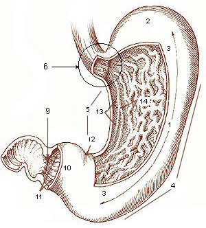
Macroscopic structure of the stomach. (1) corpus, (2) fundus, (3) anterior wall of stomach, (4) major curvature, (5) minor curvature, (6) cardia, (9) sphincter pylori, (10) antrum, (11) canalis pyloricus, (12) incisura angularis, (13) gastric groove, (14) mucosal folds (by fenestration).
Questions and Answers
Q: What is the stomach?
A: The stomach is part of the digestive system. It is an internal organ between the esophagus and small intestines.
Q: What is the function of hydrochloric acid in the stomach?
A: Hydrochloric acid made by stomach cells has a few functions. As a useful side-effect, it kills bacteria in food, but its main purpose is to help protein-digesting enzymes called proteases work.
Q: How does food move through the digestive process?
A: Food moves through the digestive process by being held in the stomach after ingestion, then passing into the small intestine where most of its nutrition is absorbed. Partially digested food (chyme) then passes out of the stomach and into other parts of digestion.
Q: What are some problems that can occur with too much stomach acid?
A: Too much stomach acid can make peptic ulcers worse and cause heartburn—pain in chest when acid from stomach refluxes back up into oesophagus. These can usually be treated by neutralizing the acid. Vomiting may also occur when food goes back up oesophagus and out mouth, which can be a symptom of disease.
Q: How do ruminants' digestive systems differ from humans'?
A: Ruminants have a multi-compartmented stomach where food goes through first part of digestion twice, and digesting grass is done by bacteria instead of human digestion processes like churning, squeezing and mixing for preparation for digestion as seen in humans.
Q: Are there any other words for "stomach"?
A: Yes, other words for "stomach" include belly and tummy
Search within the encyclopedia