Retina: structure, function, and clinical importance
The retina is the light-sensitive neural layer lining the back of the eye. This article explains its anatomy, photoreceptors, signal pathways, clinical conditions, imaging, and variation across species.
Overview
The retina is the thin, multi-layered neural tissue that lines the inner surface of the eyeball. It detects incoming light and begins the conversion of photons into electrical signals. These signals are processed locally in retinal circuits and then transmitted to the brain so that visual scenes can be recognized and used to guide behavior.
Image gallery
10 Images


Structure and main parts
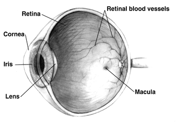
Histologically the retina contains several principal layers: the outer retinal pigment epithelium, the photoreceptor layer (rods and cones), interneurons (bipolar, horizontal, and amacrine cells), and the ganglion cell layer whose axons form the optic nerve. Photoreceptors house light-sensitive pigments (opsins) and perform phototransduction. The central region specialized for high-acuity vision is the macula; at its center lies the fovea, a small pit densely packed with cones and largely devoid of rods.
Photoreceptors and function
There are two main photoreceptor types: rods are highly sensitive and support dim-light (scotopic) vision, while cones mediate color and high-resolution (photopic) vision. Phototransduction converts light into ionic changes inside photoreceptors, creating signals that traverse retinal interneurons to retinal ganglion cells. Many retinal neurons are true neurons that perform substantial preprocessing before information leaves the eye.
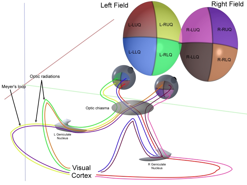
Pathways to the brain and non-image functions
Most retinal output travels in the optic nerve to visual centers for image formation. A distinct set of intrinsically photosensitive retinal ganglion cells provides input via the retino-hypothalamic tract to brain structures that regulate circadian rhythms and pupil responses. Thus the retina contributes both to perception and to physiological adjustments tied to the 24-hour day.
Clinical relevance and imaging
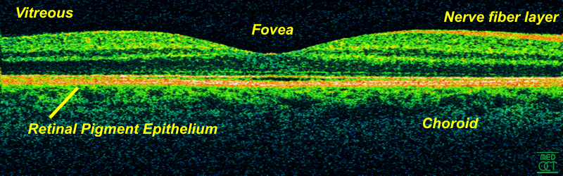
Retinal disease is a major cause of visual loss. Common conditions include age-related macular degeneration, diabetic retinopathy, retinal detachment, and inherited retinopathies such as retinitis pigmentosa. Modern clinical tools — direct and indirect ophthalmoscopy, fundus photography, and optical coherence tomography — allow detailed visualization of retinal layers and guide treatment.
Comparative and notable facts
Across species the retina adapts to ecological needs: nocturnal animals have rod-dominant retinas, many birds and some fish have additional cone types for expanded color vision, and some mammals possess a reflective layer (tapetum) to enhance sensitivity. Developmentally the retina arises from the neural tube as an outpouching of the brain, underscoring its identity as central nervous system tissue. For general introductions and further reading see brain-vision links, basic texts on visual perception, and clinical resources cited in specialist sources such as eye anatomy references.
- Key components: photoreceptors, pigment epithelium, bipolar and ganglion cells.
- Common tests: visual acuity, fundoscopy, electroretinography, imaging.
- Where to learn more: specialist ophthalmology texts and review articles on retinal disease and on photoreceptor biology.
Retinal research remains active, spanning molecular studies of phototransduction, regenerative approaches, gene therapies for inherited disorders, and prosthetic interfaces that aim to restore sight. For authoritative clinical guidelines and research updates consult professional ophthalmology organizations and peer-reviewed literature indexed through standard medical libraries (visual science resources, clinical guidelines, imaging protocols).
Related articles
Author
AlegsaOnline.com Retina: structure, function, and clinical importance Leandro Alegsa
URL: https://en.alegsaonline.com/art/82345