
Visual cortex: structure, function, development and methods of study
The visual cortex is the occipital brain region that processes visual information. This article summarizes its anatomy, functional areas, development, common research methods, and clinical significance.
Overview
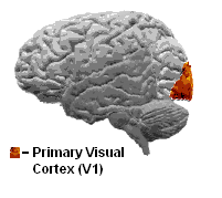
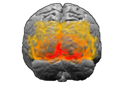
The visual cortex is the part of the cerebral cortex that receives and interprets visual signals. Located in the occipital lobe at the back of the brain, it transforms inputs from the retina (via the lateral geniculate nucleus) into representations of edges, motion, color and spatial relationships. The primary visual cortex, often called V1, is thin in humans (roughly 1.5–2 mm) but occupies a large fraction of cortex in many primates and other mammals. For general context on brain anatomy see brain overview and for introductory material on vision see vision science.
Image gallery
8 Images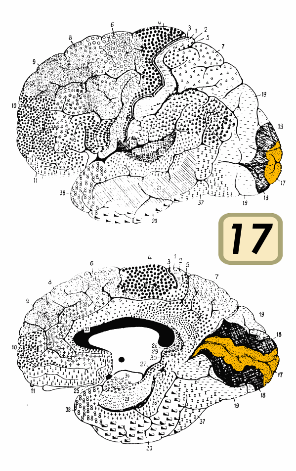




Structure and organization
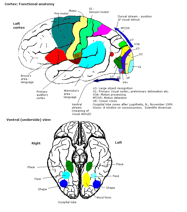
The visual cortex is arranged into multiple areas with distinct roles. V1 (primary visual cortex) is the main cortical input site; subsequent extrastriate areas include V2, V3, V4 and MT/V5, which contribute to form, color and motion processing. V1 itself has a layered, six-layer architecture and contains functional modules such as orientation columns and ocular dominance columns that reflect how neurons respond to line orientation and to inputs from one eye or the other. The mapping from the retina to cortex is retinotopic, with central vision occupying disproportionate cortical territory (cortical magnification). Basic anatomy and layering are discussed in resources such as cortical layers and primates' cortex.
Functions and examples
Neurons in the visual cortex perform elementary computations that underlie perception: detecting edges and orientations, signaling direction and speed of motion, discriminating color, and combining inputs for stereoscopic depth. These detectors are the building blocks that higher areas use to recognize objects and guide visually guided actions. The cortex participates in both the ventral "what" stream (object recognition) and the dorsal "where/how" stream (motion, spatial relationships). For applied or comparative examples see comparative vision and occipital lobe function.
Development, plasticity and notable discoveries
Development of visual cortical circuits depends on patterned visual experience during critical periods early in life. Classic experiments by David Hubel and Torsten Wiesel revealed how deprivation or altered input during development changes ocular dominance and receptive-field properties; their work earned the 1981 Nobel Prize for contributions to understanding sensory processing and plasticity. For historical and biographical notes see Hubel biography and Wiesel biography. Their studies linked single-neuron responses to emergent properties such as edge detection and stereoscopic depth (Nobel context).
Research methods
Investigating the visual cortex uses a range of techniques. Invasive single-unit and multi-electrode recordings measure action potentials directly in animal models such as cats, ferrets, rats, mice and monkeys; see experimental approaches in cat studies, ferret studies, rat studies, mouse studies and monkey studies. Noninvasive human techniques include electroencephalography (EEG), magnetoencephalography (MEG) and functional magnetic resonance imaging (fMRI), which track ensemble activity and blood-flow correlates of neural processing; see methodological summaries at action potentials, electrode methods, and EEG resources as well as fMRI basics. Additional overviews of sensory signaling pathways are available at visual signal pathways.
Clinical relevance and distinctions
- Cortical blindness: damage to primary visual cortex can produce loss of conscious vision despite intact eyes; some patients show residual nonconscious abilities known as blindsight.
- Plasticity and rehabilitation: limited recovery of function can occur after injury, and therapies sometimes target cortical remapping.
- Species differences: the relative size and organization of visual cortex vary widely across mammals; primates generally have larger, more elaborated visual cortical areas.
For further reading and curated resources on anatomy, physiology and clinical aspects consult introductory and specialist material such as neuroanatomy guides, reviews on cortical processing (vision reviews), and experimental technique primers (electrophysiology, EEG, fMRI). More targeted discussions of cortical modules, developmental plasticity and comparative neuroanatomy are available through the linked resources: comparative studies, historical summaries, and research archives.
Questions and answers
Q: What is the visual cortex?
A: The visual cortex is a part of the brain that allows vision. It is located in the occipital lobe at the back of the brain and is relatively thin, between 1.5mm and 2mm in humans.
Q: Who did research on the visual cortex?
A: David Hubel and Torsten Wiesel did research on the visual cortex for many years. They won the 1981 Nobel Prize in Physiology or Medicine for their discoveries about information processing in the visual system.
Q: What kind of research did they do?
A: Their work in the 1960s and 1970s was on how the visual system developed. They worked on parts of the visual cortex of the brain which get signals from either eye, describing how signals from these eyes are processed by the brain to generate edge detectors, motion detectors, stereoscopic depth detectors and colour detectors - building blocks of a visual scene.
Q: How can researchers study primary visual cortex activity?
A: Research on primary visual cortex activity can involve recording action potentials from electrodes within an animal's brain (cats, ferrets, rats, mice or monkeys). Alternatively, signals can be recorded outside an animal using EEG, MEG or fMRI techniques which gather information without invading its brain.
Q: How thick is human's Visual Cortex?
A: Human's Visual Cortex is relatively thin - between 1.5mm and 2mm thick.
Q: What award did Hubel and Wiesel win for their discoveries about information processing in Visual System?
A: David Hubel and Torsten Wiesel won 1981 Nobel Prize in Physiology or Medicine for their discoveries about information processing in Visual System
Related articles
Author
AlegsaOnline.com Visual cortex: structure, function, development and methods of study Leandro Alegsa
URL: https://en.alegsaonline.com/art/105637
Sources
- sciencedirect.com : sciencedirect.com/science/article/pii/S0042698910003743