Cone cell (photoreceptor): structure, function and role in color vision
Cone cells are retinal photoreceptors that mediate daytime vision, high spatial acuity and color discrimination. This article covers their anatomy, physiology, spectral types, distribution and clinical relevance.
Overview
Cone cells, or cones, are specialized photoreceptor neurons in the vertebrate retina that transduce incoming light into electrical signals used by the visual system. Cones function best under well-lit (photopic) conditions and are primarily responsible for color perception, fine detail and rapid temporal resolution.
Image gallery
2 Images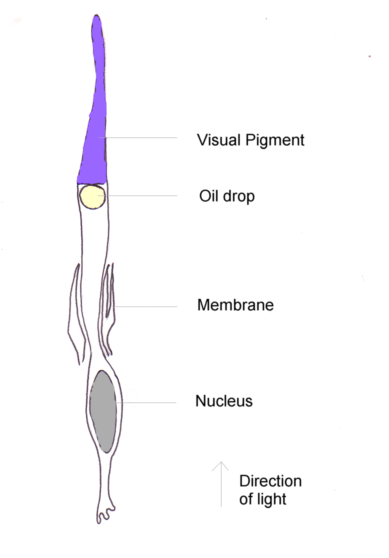
Structure and physiology
Each cone has an outer segment containing stacks of membranous disks or folds where light-sensitive pigment molecules reside, an inner segment with mitochondria and synthetic machinery, a cell body and a synaptic terminal that transmits signals to bipolar and horizontal cells. Phototransduction in cones uses a G-protein cascade similar to rods but with molecular differences that reduce amplification and speed recovery, allowing cones to respond and adapt quickly to changing light levels. Cones generally have faster responses than rods and contribute to higher temporal and spatial resolution.
Spectral types and opsins
Human retinas typically contain three classes of cones, distinguished by the opsin protein they express in the outer segment. These opsins, sometimes called photopsins, have peak sensitivities at short (S), medium (M) and long (L) wavelengths, supporting trichromatic color vision. The relative stimulation of these cone types is compared by retinal and cortical circuits to encode hue and saturation. Some animals possess four or more cone classes, enabling tetrachromacy or broader spectral discrimination.
Distribution in the retina
Cones are unevenly distributed: a very high density occupies the fovea centralis, the retinal region for the sharpest central vision, while their density falls toward the periphery where rods predominate. This distribution underlies the difference between high-acuity, color-rich central vision and more light-sensitive peripheral vision. Cones also adapt their sensitivity through pigment bleaching and neural mechanisms, allowing useful function across a range of daylight intensities.
Comparison with rods
- Light sensitivity: rod cells are more sensitive in dim light but do not encode color.
- Temporal and spatial resolution: cones provide faster responses and greater acuity.
- Functional roles: cones dominate daytime tasks such as reading and color discrimination, while rods support scotopic (low-light) vision.
Development, variation and clinical significance
Cone number and distribution are established during retinal development and can vary between individuals. Early histological estimates, such as Osterberg's work, suggested on the order of millions of cones per human eye; modern imaging refines those counts and shows individual variability. Genetic or acquired defects affecting cone structure or opsins cause clinical conditions including inherited color vision deficiencies, achromatopsia and cone dystrophies. Terms such as color blind describe a range of color-perception deficits resulting from absence or alteration of one or more cone types.
Testing and diagnosis
Clinical tests assess cone function and color discrimination. Psychophysical color tests, ophthalmoscopy and retinal imaging evaluate cone health and distribution, while electroretinography can measure cone-driven electrical responses. Genetic testing can identify mutations in opsin genes and other components relevant to cone disorders. For general background see resources on the retina and on visual physiology at introductory and clinical sites (human vision references and eye health materials).
Evolutionary and applied notes
Trichromacy and related cone specializations have evolved independently in several lineages and are thought to offer advantages for tasks such as detecting ripe fruit and social signals. Knowledge of cone spectral responses informs colorimetry, display design and lighting standards. Research into cone regeneration, gene therapies and prosthetic devices aims to restore or augment cone function in retinal disease.
Further reading
For an introduction to photoreceptor biology consult general photoreceptor overviews and texts on retinal physiology. Comparative vision discussions may be found in resources covering animal photoreceptors and evolutionary ecology, and molecular details appear in works on photopsins and opsin genetics. Clinical and diagnostic guidance is available through ophthalmology references and genetic counseling materials (visual physiology), while historical population studies and reviews discuss counts and distributions of cones (rod vs cone material and population surveys).
Readers seeking patient-oriented information should consult reputable eye health sources and color vision testing guides. For technical protocols and current research summaries, see specialist reviews and laboratory resources in visual neuroscience and molecular photobiology (experimental methods and color vision literature).
Related articles
Author
AlegsaOnline.com Cone cell (photoreceptor): structure, function and role in color vision Leandro Alegsa
URL: https://en.alegsaonline.com/art/22454