Midbrain (Mesencephalon): Structure, Function, and Clinical Significance
A concise encyclopedia entry on the midbrain (mesencephalon): its anatomy, major nuclei and pathways, development, roles in sensation and motor control, evolution, and clinical importance.
Overview
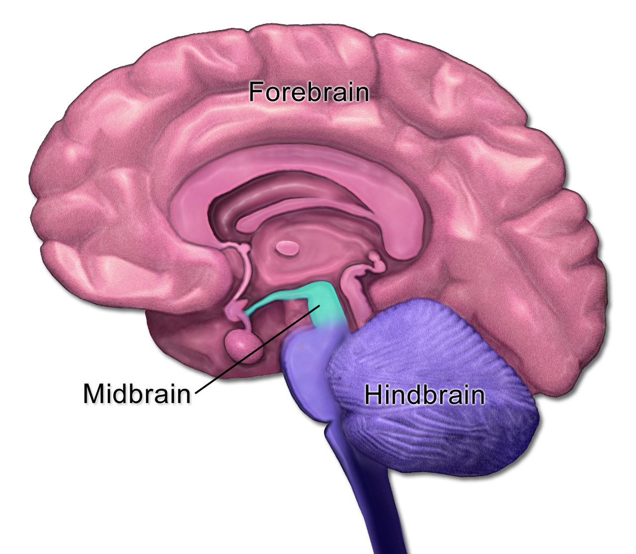
The midbrain, or mesencephalon, is the middle segment of the brainstem and a compact hub of neural circuits that link the forebrain with the hindbrain. It participates in sensory processing, motor control, regulation of arousal and sleep/wake states, and several reflexive behaviors. The midbrain sits above the pons and medulla and below parts of the diencephalon, forming a central conduit of ascending and descending pathways and cranial nerve connections (brain stem).
Image gallery
7 Images


Structure and major components
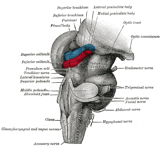
Anatomically the midbrain can be divided into a dorsal tectum and a ventral tegmentum, separated by the aqueduct of the midbrain. Key nuclei and fiber systems include:
- Tectum: superior and inferior colliculi — centers for integration of visual and auditory reflexes, respectively (vision, hearing).
- Tegmentum: contains the red nucleus, oculomotor (III) and trochlear (IV) nerve nuclei, and reticular formation important for motor coordination and arousal.
- Substantia nigra and ventral tegmental area: dopaminergic nuclei that influence movement, motivation and reward processing (dopamine).
Development and evolutionary context
During embryonic development the central nervous system forms from the neural tube, which gives rise to three primary vesicles; the mesencephalon corresponds to the middle vesicle and later becomes the midbrain segment of the brain. Classic descriptions of vertebrate brain development identify the paired and regionalized vesicles that shape mature anatomy (vesicles, anatomy). The midbrain is considered phylogenetically ancient: its basic organization and many circuits are conserved across distant vertebrate groups (architecture, vertebrates), reflecting core roles in orienting responses and sensorimotor integration.
Functions and clinical relevance
The midbrain supports a range of functions by routing and transforming sensory signals and by issuing motor commands. Important roles include:
- Reflexive orientation to visual and auditory cues via the superior and inferior colliculi.
- Control of eye movements and pupillary responses through oculomotor nuclei.
- Modulation of movement and posture through dopaminergic projections from the substantia nigra; disruption of this system is central to movement disorders such as Parkinsonian syndromes.
- Contributions to attention, arousal and aspects of reward and motivation linked to midbrain dopamine neurons.
Because of these roles, lesions or degeneration affecting midbrain structures can produce characteristic syndromes: parkinsonism, impaired vertical gaze, pupillary abnormalities, and disrupted consciousness. The midbrain’s neurotransmitter systems are not only relevant for human clinical neurology but also for comparative studies: dopamine from midbrain sources influences learning and habituation across many animal species, a function that has parallels even outside vertebrates (habituation, insects).
Notable distinctions
The midbrain should be distinguished from adjacent forebrain structures that handle higher-order sensory analysis and from hindbrain regions more specialized for basic life support. Its compact size belies the diversity of nuclei it contains and the number of ascending and descending tracts that pass through it, making it a strategic bottleneck in both normal function and disease.
For further anatomical diagrams and functional maps consult specialized resources and neuroanatomy references (brain stem, anatomy, vision, hearing, brain, vesicles, architecture, vertebrates, dopamine, habituation, insects).
Related articles
Author
AlegsaOnline.com Midbrain (Mesencephalon): Structure, Function, and Clinical Significance Leandro Alegsa
URL: https://en.alegsaonline.com/art/64655