
Brain stem: structure, functions, and clinical importance
Overview of the brain stem—its anatomy (midbrain, pons, medulla), core functions in autonomic control and cranial nerves, clinical significance, development, blood supply, and distinguishing features.
Overview
The brain stem is the lower, posterior continuation of the brain that physically and functionally links the cerebrum and cerebellum to the spinal cord. Though comparatively small, it houses networks essential for life-sustaining processes and serves as a conduit for the major motor and sensory pathways between the brain and the body.
Image gallery
9 Images




Anatomy and main components
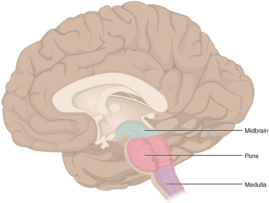
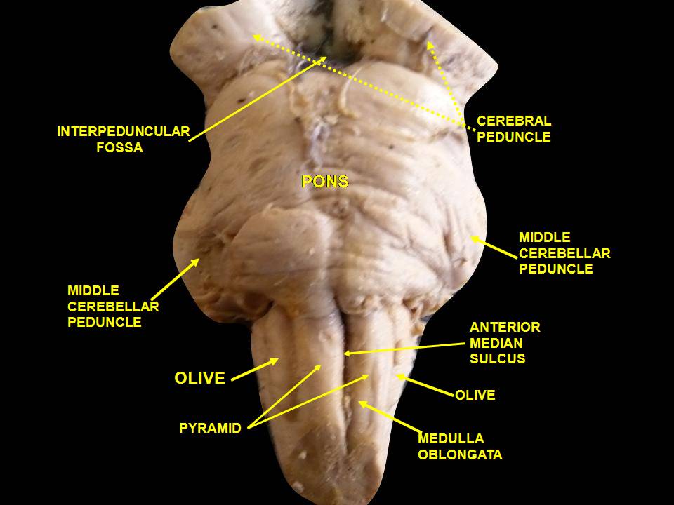
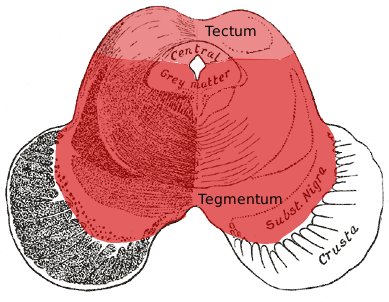
Anatomically the brain stem is commonly divided into three regions. The midbrain sits superiorly; beneath it lies the pons; and caudally the medulla oblongata. The medulla is continuous with the spinal cord. The hindbrain grouping often used in older classifications includes both the pons and medulla and is sometimes contrasted with the midbrain as the hindbrain.
Functional components
Within these regions are multiple specialized nuclei and axonal tracts. The brain stem contains origin nuclei for most of the cranial nerves and therefore gives rise to the principal motor and sensory nerves of the head and neck; for example, many fibers of the cranial nerves emerge there and serve structures such as the face. Long ascending sensory and descending motor pathways transit the brain stem, providing the main route from the cerebral hemispheres and cerebellum to the rest of the body (and vice versa).
Key roles and regulation
Core involuntary functions are coordinated in the brain stem. Embedded neuronal groups and networks regulate breathing, cardiovascular reflexes, and basic arousal states, as well as autonomic responses like sweating. The neurons that mediate these reflexes and rhythms include brain stem neurons and associated circuits. In particular, centers in the medulla help control respiration and elements that influence blood pressure. The reticular formation, a diffuse set of nuclei that runs through the brain stem, contributes to wakefulness and attention.
Clinical significance
Because the brain stem contains vital centers and the main conduits for nerve signals, injury, stroke, or compression here can produce severe and sometimes life-threatening deficits. Damage may impair breathing and heart rate, alter consciousness, or produce characteristic syndromes affecting cranial nerve function plus motor or sensory pathways. Understanding which motor and sensory tracts and cranial nerve nuclei lie at particular levels of the brain stem helps clinicians localize lesions and predict outcomes.
Development, blood supply, and distinctions
During embryonic development the midbrain and hindbrain arise from distinct segments of the early neural tube; the hindbrain then differentiates to form structures that integrate with the cerebrum and cerebellum. The brain stem's blood supply is principally provided by branches of the vertebral and basilar arterial systems; disruption of these vessels can produce characteristic infarcts. Compared with the bulkier cerebral hemispheres, the brain stem is compact but densely organized, containing many nuclei and pathways in only a few centimeters of tissue.
- Major parts: midbrain, pons, medulla oblongata.
- Connections: links to spinal cord, cerebrum, and cerebellum.
- Functions: autonomic control, cranial nerve origins, conduction of ascending and descending pathways, arousal and reflexes.
- Clinical notes: lesions affect breathing, circulation, consciousness, and cranial nerve function; rapid diagnosis is critical.
For an illustrated orientation to brain stem anatomy and its clinical correlations, consult neuroanatomy resources or trusted clinical references (see further reading). Additional general material is available in standard neurology texts and anatomical atlases (overview, hindbrain, cranial structures).
For targeted study, review the organization of cranial nerve nuclei, the pathways that ascend to the thalamus and descend from the cortex, and the vascular territories supplied by vertebrobasilar arteries. Those topics clarify why small lesions in the compact brain stem can produce complex, multisystem deficits.
Questions and answers
Q: What is the brainstem?
A: The brainstem is the rear part of the brain.
Q: What are the two sections of the brainstem?
A: The two sections of the brainstem are the hindbrain and the midbrain.
Q: What does the motor and sensory connections pass through in the body?
A: The motor and sensory connections from the main part of the brain to the rest of the body pass through the brain stem.
Q: What are the cranial nerves?
A: The cranial nerves are the main motor and sensory nerves to the face and neck.
Q: What functions does the brainstem control?
A: The brainstem controls bodily functions that are necessary for life, such as breathing, heart beat, and sweating.
Q: What are the two components of the hindbrain?
A: The two components of the hindbrain are the pons and medulla oblongata.
Q: What does the medulla oblongata do?
A: The medulla oblongata, which is an extension of the spinal cord, is the control center of bodily functions such as breathing and blood pressure.
Related articles
Author
AlegsaOnline.com Brain stem: structure, functions, and clinical importance Leandro Alegsa
URL: https://en.alegsaonline.com/art/13658