Insect
Insects (Insecta), also known as notch animals, are the most species-rich class of arthropods and at the same time the most species-rich class of animals of all. Almost one million insect species have been scientifically described to date (925,000 according to Grimaldi/Engel 2005, 865,000 according to Nielsen/Mound 1997). This means that more than 60 percent of all described animal species are insects. According to various extrapolations, however, it is estimated that the figure is many times higher, with millions of undiscovered species still suspected, especially in the tropical rainforests. The first fossil record of insects dates back to the Devonian period, about 400 million years ago.
The word "insect" (from Latin īnsectum "incised") was adopted as a foreign word in the 18th century and thus means "incised (animal)", referring to the strongly separated body parts. It is a loan translation of Ancient Greek ἔντομα [ζῶα] éntoma zōa, German 'insects, notched animals' (literally 'incised [animals]'; to ἐντέμνειν entémnein, German 'to incise'), which is included in entomology (entomology). The term 'Kerbtier' goes back to the German writer Philipp von Zesen. In the past, the scientific name Hexapoda (Greek for 'six-footed') was also used, which is now reserved for a higher-level group (see systematics section).
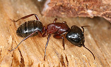
Ants, like the brown-black horse ant here, show the insects' body parts, which are separated from each other, particularly clearly.
External anatomy
The size of the insects varies greatly, ranging from one to 20 millimetres for most species. The smallest species are hymenoptera (dwarf wasps) parasitizing in eggs of other insects, the male of the smallest known species is 0.15 (to 0.24) mm long. The smallest free-living insects are dwarf beetles with a body length of 0.4 mm (even smaller figures are based on measurement errors). The largest known insects are stick insects with a body length of about 33 centimetres and the giant longhorn beetle (Titanus giganteus) with a body length of 15 centimetres. According to their way of life, the body of the insects can be elongated, flattened or more or less spherical.
Common to all insects is an exoskeleton made of the solidifying structural protein sclerotin and the polysaccharide chitin, as well as the anatomically usually clearly visible division into the three sections (tagmata) head (caput), thorax and abdomen and the presence of exactly three pairs of legs. All body parts consist of individual segments, which have a dorsal plate (tergite), an abdominal plate (sternite) and lateral plates (pleurites). On the sides of the individual segments, openings of the tracheal system, so-called stigmas, are laid out in the basic structural plan, but their actual number can vary greatly among the individual taxa of insects. Extremities and their parts (mouthparts, styli, gonopods) may also occur in pairs on each individual segment. The thorax bears six legs (three pairs). Each segment also has a pair of ganglia in the abdominal medulla, with the ganglia of the head fused to form an upper-limb ganglion and a lower-limb ganglion. Between each segment are stretchable membranes, the intersegmental membranes, which allow the segments to move against each other and the body to change volume during egg production, feeding, or respiration. In rigidly fused segments, such as in the head, these cuticles are not present.
The exoskeleton forms the outer layer of the insect body. This cuticle is formed by an underlying epidermis. Embedded in it are the sensory organs and various glandular outlets of the insect. With more or less involvement of the epidermis, the exoskeleton forms various surface structures, which include warts, spines, hairs, bristles, scales and humps. The outer layer of the insect or individual parts of the body can be coloured due to the incorporation of pigments or due to special light-refracting surfaces (interference colours).
The sensory organs are mainly hair sensilla, which are distributed over the body. These react to vibrations and oscillations, but can also perceive odours, moisture or temperatures. Some of these sensory cells are grouped into sensory organs, such as Johnston's organ on the pedicellus of the antenna or the tympanic organs for sound perception, which are found, for example, in the long-sensed grasshoppers. This allows the perception of vibrations in the range of 1 Hz to 100,000 Hz. The compound eyes and the ocelli serve as optical sense organs, in many larvae also the larval dot eyes.
The insect head: eyes, antennae, mouth parts
The insect head consists of an anterior segment, the segmental nature of which is disputed (according to the previously prevailing Articulata hypothesis, it corresponds to the acron of the annelids) and five further fused segments and bears the eyes and the limb antenna as well as a typical apparatus of mouthparts consisting of paired mandibles and maxillae as well as an unpaired labium. Accordingly, the mouth opening, through which food is taken in, is also located on the head. The externally visible sutures of the insect head have nothing to do with the segment boundaries except for one exception, the boundary between the fifth and sixth segment (postoccipital suture).
The compound eyes are located on the anterior segment, besides which insects possess three point eyes (ocelli) in the basic structural plan for light-dark perception. The second segment is the antennal segment with the flagellar antenna typical for insects. This differs from the limb antenna found in the original hexapods in that it has only one basal or shaft limb with musculature, the scapus. The following turning limb, the pedicellus, is movable together with the flagellum in relation to the scapus. The pedicellus contains Johnston's organ, a sensory organ that can respond to vibration and sound. The flagellum itself varies in length and can also be very differently shaped. It has sensory cells that serve primarily to perceive smell.
The third segment is the intercalary segment, which again has no structures of its own (it corresponds to the second antennal segment of crustaceans). At the fourth to sixth segment are the mouthparts, beginning with the mandibles and the maxillae, each of which are present in pairs, and concluding with the labium, which is unpaired due to an adhesion of the basal phalanx and closes the oral cavity to the rear. The anterior termination is formed by the clypeus with the labrum. The mandible consists of only one limb. As a biting and chewing tool, it represents the most powerful mouth tool in most insects. The maxilla shows the extremity character of the mouthparts very clearly and reminds of the split bone of crustaceans. It consists of a basal limb, the coxopodite, divided by a cardo and a stipes. This is followed by two caulades, called the galea and lacinia, and a multimembered palpus, the palpus maxillaris. The labium corresponds to this structure, with the exception that here the two coxopodites are fused and form a common basal plate, which is divided into mentum and submentum. Here, too, there are two caulæ, the glossae and the paraglossae, on each side, and a palpus, the palpus labialis. Especially the palps have sensory cells for the perception of smell and taste. Further structures of the oral cavity are the hypopharynx as a tongue-shaped formation of the oral vestibule and the epipharynx on the inner side of the labrum.
The construction plan described here corresponds to the basic pattern of insects and is called the chewing-biting type. Through modifications of the individual structures in adaptation to different diets, various other types of mouthparts can develop from this type, including the licking-sucking mouthparts of many flies or the biting-sucking mouthparts of mosquitoes or bugs.
The thorax
The thorax always consists of three segments and carries the six walking legs in all insects as well as the two pairs of wings in winged insects. The individual segments are called prothorax, mesothorax and metathorax because of their position relative to each other. Each of these segments bears a pair of thoracic legs, which are commonly pronounced as walking legs. These legs can vary greatly in shape, depending on their function, but always have the same basic structure. They consist of a hip (coxa), a following thigh ring (trochanter), the thigh (femur), the splint (tibia) as well as a jointed foot (tarsus), which consists of one to five tarsal limbs as well as a praetarsus. The praetarsus normally bears two claws and, in many insects, several other structures that serve primarily to hold on to various surfaces.
In winged insects, the meso- and metathorax also each bear a pair of wings, which can be differently pronounced in the various insect groups. Since the meso- and metathorax form a unit here, they are also referred to together as the pterothorax (from Greek pteron = "wing, feather, fin").
However, the original insect groups such as the rock jumpers and the fishies are unwinged; accordingly, wings are not present in the basic structural plan of insects and only developed later within insects, with the evolution of flying insects. Instead, insects in the basic structural plan have duplications of the epidermis laterally on the thoracic segments, which are called paranota and are considered to be precursor structures of the wings.
The abdomen
The abdomen originally consists of eleven segments, of which, however, some have been fused or lost in the various insect taxa. The abdominal segments bear only modified extremities and no real legs. Styli, for instance, are found, especially in larval forms. In addition, a number of insect groups possess cerci on the last segment, which are formed for different functions. The gonopods, which form the mating organs of males and the ovipositor of females, are also transformed extremities. In contrast to the thorax, the abdomen contains very little musculature; instead, most organ systems are located here.
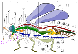
Scheme of insect anatomyA - Caput (head) B - Thorax (chest) C - Abdomen (abdomen). 1. antenna2 . Ocellus (front) 3. Ocellus (top) 4. Complex eye (compound eye) 5. Brain (superior ganglion) 6. Prothorax7 . Posterior (dorsal) artery8 . Trachea9 . Mesothorax10 . Metathorax11 . First pair of wings12 . Second pair of wings13 . Midgut14 . heart15. ovary16 . Hindgut (rectum) 17. anus18 . Vagina19. ventral nervous system with ganglia20 . Malpighian vessels21 . Tarsomere22 . Praetarsus23 . Tarsus24 . Tibia25 . femur26 . Trochanter27 . Foregut28. thoracic ganglion29 . Coxa30 . Salivary gland31 . Submandibular ganglion32 . Mouthparts
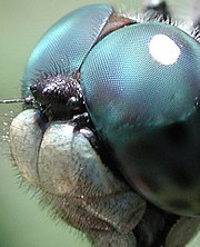
Head of a dragonfly with compound eye and ocelli
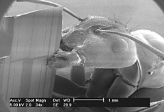
Harvester ant in the scanning electron microscope
.jpg)
Diced dance fly (Empis tesselata)
.jpg)
pubescence of a bumble bee
Internal Anatomy and Physiology
Nervous and endocrine system
The nervous system of insects essentially corresponds to the common basic structural plan of the Mandibulata, to which crustaceans and millipedes also belong. It consists of a paired abdominal cord, which has a pair of ganglia in each segment. In the head, the ganglia are fused to form an upper pharyngeal ganglion, the brain, as well as a lower pharyngeal ganglion, both of which are connected to each other by the pharyngeal connectives. The brain itself has a very large anterior area, the protocerebrum, from which areas radiate bilaterally into the centers of the compound eyes, called lobi optici. The ocelli are also innervated by this part of the brain. From the deuterocerebrum, the second brain section, nerves extend into the antennae, and the tritocerebrum supplies nerves to the foregut through a tritocerebral commissure. The mouthparts are supplied by the ganglia of the fourth to sixth segments concentrated in the submandibular ganglion. The abdominal medulla runs through the thorax in the form of a typical rope ladder nervous system, with three particularly large pairs of ganglia in the thorax supplying the legs and (when present) the wings. The abdomen contains 7 normal pairs of ganglia in the basic structural plan and a ganglion mass in the eighth abdominal segment, which consists of the ganglia of all the following segments.
The autonomic nervous system consists of three sections. The anterior section is formed by the nerves of the oral and foregut region with the frontal, hypocerebral and ventricular ganglion as well as the corpora cardiaca and the corpora allata. The second section is formed by the paired abdominal ganglion chain, which is responsible for innervation of the stigmata. The posterior intestinal segment and the genitalia are supplied by the caudal nerve. Particularly in the brain, insects also possess neurosecretory glands, which include the Corpora cardiaca and Corpora allata, already named. The latter secrete the juvenile hormone, which influences the developmental stage during moulting. The moult itself is induced by the moulting hormones, especially the ecdysone.
Breathe
The respiratory organs of insects are largely rigid tubes that run through the entire body and extend to the organs and individual cells in increasingly fine tubes. This system is called the tracheal system, and develops from invaginations of the epidermis towards the interior. Accordingly, like the outside of insects, the tracheae are covered with an epidermis and a lining of chitin. This cuticle is stiffened by taenidia, spiral structures that wrap around the tube to prevent collapse. Outwardly, the tracheae open into respiratory openings called stigmas.
The breathing process is primarily passive. This form of breathing is also the reason why insects can grow larger in warmer areas. In rather cold areas, the molecular movement of the air is not sufficient to supply the internal tracheae with sufficient oxygen. However, air exchange is regulated by muscular control of the stigmas and can be increased by ventilation. In many insects the number of stigmas has been reduced from the original one pair on each thoracic and abdominal segment by cross-connections (anastomoses), and air sacs have also evolved in many taxa to aid respiration and store oxygen. In aquatic insects, additional adaptations have also evolved to allow respiration underwater. For example, many aquatic beetles and other aquatic insects are found to have an area with a special surface that allows them to hold an air bladder (physical gill). Other species have a constant volume plastron or breathing tubes for respiration. Especially in aquatic insect larvae, tracheal respiration is completely abandoned and oxygen is instead taken in through tracheal gills or the skin.
Circulation
The blood vessel system of insects essentially consists of a cavity, the haemocoel, in which all internal organs swim in blood (haemolymph) (open blood circulation).
As the remaining part of the vascular system, insects possess an unbranched, simple dorsal vessel. This has a contractile part in the abdomen, which pumps the haemolymph into the body and is accordingly called the heart (closed caudally), and a branch which supplies blood to the head, the brain and the head and is called the aorta (open cranially). The flow of blood is kept in motion by the peristalsis of the heart, haemolymph being drawn into the heart from one to twelve pairs of lateral openings of the heart, the ostia, and pumped forward through the aorta into the head. From there it distributes through the thorax into the abdomen and also into the legs and wings of the animal. On the legs, antennae, and wings, smaller pumping systems provide a supply to the ends of the structures. Thus in the head there is a muscular so-called antennal heart, it pumps the haemolymph into the antennal ampullae and into the associated antennae. Below the heart lies a dorsal membrane of musculature and connective tissue, which acts as a diaphragm separating the dorsal part of the body (pericardial sinus) from the main space of the body (perivisceral sinus) containing the intestine and the reproductive organs. Another diaphragm lies above the abdominal part (perineuralsinus) with the abdominal marrow.
See also: "Heart types and their distribution in the animal kingdom" in the article Heart
The hemolymph itself makes up 20 to 40 percent of the body's weight and consists of cells called hemocytes, as well as fluid plasma. It transports the carbon dioxide produced during respiration, proteins, nutrients, hormones and excretions, and also serves to regulate osmoregulation and maintain internal body pressure. The blood is usually colorless, but it can also be greenish-yellow; it not only transports nutrients to all parts of the body, but also carries hormones to their destination. The blood contains special cells that fight pathogens and others that repair damage or wounds. Unlike vertebrate blood, insect blood does not contain cells involved in respiration.
The traditional view that insect blood had lost its function of oxygen distribution due to the effective tracheal system has been relativized in recent years due to new findings. Occurrences of the red blood pigment haemoglobin have long been known in aquatic insect larvae that live under oxygen-deficient conditions, such as chironomid larvae. Genome analyses have now shown that haemoglobin production is part of the basic capability of almost all insects. Hemoglobin is predominantly found in gills and other respiratory organs, thus ensuring its function for respiration. In numerous insect orders, the blue blood pigment hemocyanin has also been detected, and at least for a stonefly, a function for oxygen supply has also been demonstrated here.
Digestion and excretion
Digestion takes place in the intestinal system of animals. This is divided into three functional sections, which are called foregut, midgut and rectum according to their position. The anterior and posterior portions are ectodermally formed and have a lining of chitin cuticle, which must also be skinned accordingly. The foregut begins with the mouth opening and consists mainly of the muscular pharynx. This section is connected to the midgut via the oesophagus, although in many insects this may also contain areas for food storage (ingluvies) or a forestomach (proventriculus) with chewing structures such as groins of chitin. The midgut is lined with a glandular epithelium and produces the enzymes necessary for digestion. This is also where the absorption of nutrients occurs, which in many insects takes place in blind tubes (caeca) or crypts. In many insects, endosymbiotic microorganisms (bacteria, fungi or flagellates) can also live in the caeca and crypts, which are required for the breakdown of certain food components, e.g. for the breakdown of cellulose. In addition, endogenous cellulase has been found in a few insects (in the termite species Reticulitermes speratus and Coptotermes formosanus). Origin of the animal cellulase gene is seen in the last common ancestor of the Bilateria.
The indigestible remains (excrement) are excreted via the rectum. The excretion of the insects takes place via small blind tubes that open into the intestine at the transition of the midgut to the rectum. These are called Malpighian vessels and, like the rectum, are of ectodermal origin. In the cells of these tubes nitrogenous excreta are actively extracted from the haemolymph and excreted with the excreta. In the rectal papillae, water is still extracted from the excretory products before excretion.
Important structures of nutrient and excreta storage are the fat bodies, which are located as large lobes in the abdomen of insects. In addition to storage, they serve for the synthesis of fats and glycogen as well as the degradation of amino acids.
Sex organs
Except for a few species, all insects are separately sexual. Very few species are hermaphrodites, a number of species reproduce by parthenogenesis.
Males possess paired testes for sperm production in the abdomen, which are connected to paired seminal vesicles (vasa seminales) via vasa deferentia. These open into an unpaired or paired ejaculatory duct, the Ductus ejaculatorius, and then through more or less complex mating organs, the Aedeagus, usually in the ninth abdominal segment to the outside. In addition, there may be supplementary glands that produce seminal fluids or substances for the formation of spermatophores and add them to the sperm.
The ovaries of females are also usually arranged in pairs. They usually consist of a cluster of individual ovarian strands called ovarioles. Each of these ovarioles consists of a germarium, where the eggs are produced, and a vittelarium for the production of the yolk cells. Depending on the way in which the eggs are supplied with yolk, three different forms of ovarioles are distinguished, which may occur in different insect taxa. In the first form, called panoistic ovariole, individual eggs in the vitellarium are supplied with yolk. In the meroistic-polytrophic type, each individual ovule has several nutritive cells and receives the yolk through them. In the meroistic-telotrophic ovariole, the single ovule remains connected to the germarium by a nutrient strand and receives the yolk through this pathway. In all types, follicle cells surround the growing oocytes.
The ovarioles unite and end in an unpaired vagina, which ends between the seventh and ninth abdominal segments either directly outwards or in a mating pocket, the bursa copulatrix (exception: mayflies). In the area of the vagina, almost all insects have a sperm collecting pocket, the receptaculum seminis, and various appendage glands for the production of putty substances or similar may also be present.
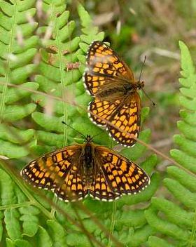
Quail Wheat Fritillary
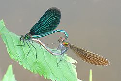
Mating of the blue-winged damselfly
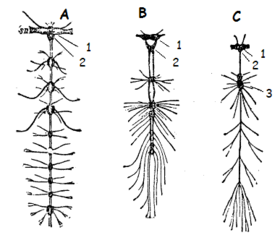
Nervous system of insects (A termite, B diving beetle, C fly) 1 superior cerebral ganglion 2 submandibular ganglion 3 other nerve node
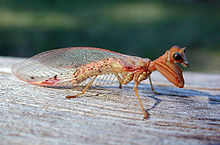
Catching from Australia: Ditaxis biseriata
Questions and Answers
Q: What is the scientific classification of insects?
A: Insects are a class in the phylum Arthropoda.
Q: How many species of insects have been identified?
A: About 926,400 different species of insects have been described.
Q: What percentage of all known living species are insects?
A: Insects make up more than half of all known living species. It is estimated that they may be over 90% of animal species on Earth.
Q: Are new insect species still being discovered?
A: Yes, new species of insects are continually being found. Estimates of the total number of species range from 2 million to 30 million.
Q: Do all adult insects have wings and six legs?
A: Yes, all adult insects have six legs; and most have wings. Insects were the first animals capable of flight.
Q: Where do most insect live?
A: Most insect live on land (terrestrial), however few live in oceans or very cold places such as Antarctica. The most species live in tropical areas.
Q: What is the name for people who study insects?
A: People who study insects are called entomologists
Search within the encyclopedia