Anatomy
![]()
The title of this article is ambiguous. For other meanings, see Anatomy (disambiguation).
Anatomy (the 'dissection' of animal and human bodies for the purpose of gaining knowledge; from the ancient Greek ἀνά aná, German 'auf', and τομή tomḗ, German 'the cutting, the section') is a branch of morphology and in medicine or human biology (anthropotomy), zoology (zootomy) and botany (phytotomy) the study of the internal structure of organisms.
The shape, position and structure of body parts, organs, tissues or cells are examined. Pathological anatomy deals with pathologically altered body parts. Microscopic anatomy deals with the finer biological structures down to the molecular level and ties in with molecular biology. Classical anatomy uses standardized nomenclature based on the Latin and Greek languages.
The term anatomy has been used since the early 16th century (also as anatomei) also more generally and transferred in the meaning "dissection, determination of structure, analysis of concrete and abstract things", also "structure, (construction)", e.g. anatomy of the soil, of art, of thought, of society.
A researcher dealing with anatomy is called an anatomist.
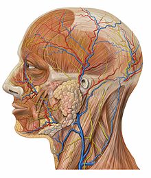
Anatomy of the human head
History
The earliest preserved anatomical studies are found in the Edwin Smith papyrus, which is dated to the 17th century BC. Among other things, the heart and coronary vessels, liver, spleen and kidneys, hypothalamus, uterus and bladder, and blood vessels are treated.
The Papyrus Ebers from the last quarter of the 16th century BC contains a treatise on the heart, in which the blood vessels are also described.
Nomenclature, methodology and applications go back to the ancient Greek physicians. Descriptions of muscles and the skeleton can be found in the Corpus Hippocraticum (especially On Fractures and On Joints), although in Hippocratic medicine human physiology was more important than anatomy. Aristotle used dissection of animals to describe the anatomy of vertebrates. Praxagoras of Kos already knew the difference between arteries and veins in the 4th century BC.
The beginnings of systematic anatomy arose in ancient Babylon. A first anatomical school existed in Alexandria in the 2nd century BC. The rulers of the Ptolemaic Empire (Ptolemy I and especially Ptolemy II come into consideration) allowed the opening of corpses there for anatomical studies, mostly on executed persons. Herophilos of Chalcedon performed the first scientific autopsies and also vivisections on humans and animals. He is said to have dissected 600 prisoners alive and is considered the "father of anatomy". He rejected Aristotle's view that the heart was the seat of the intellect and instead named the brain. Other anatomists in Alexandria were Erasistratos and Eudemos of Alexandria.
The treatise On the Designation of the Parts of the Human Body, written by Rufus of Ephesus in the 2nd century, is the oldest extant anatomical textbook whose main purpose was to teach anatomical nomenclature. According to Rufus, theoretical instruction was supplemented by illustrations on living persons, with the external body parts being demonstrated on slaves.
In the 2nd century AD, Galenos of Pergamon systematically summarized the medical knowledge of ancient physicians, among other things in a 15-volume anatomical work On the Procedure of Dissection. As a doctor of gladiators, he was able to study various types of wounds and thus also human anatomy in detail. He carried out further studies on pigs and monkeys. His writings formed the basis for the works of the Middle Ages, including Avicenna's Canon of Medicine.
From about 1300 onwards, anatomical doctrinal dissections were occasionally performed, especially in Upper Italy. Such demonstrations, however, served primarily the purpose of confirming the teachings of ancient authors or authorities.
From the 15th century onwards, anatomy experienced a new impetus, inspired by ideas of humanism and the Renaissance. After anatomy had failed to make much progress in the Middle Ages, the Flemish anatomist Andreas Vesalius (1514-1564) corrected assumptions or beliefs that had hardly been questioned for centuries, outraging many of his colleagues. His work made him the founder of modern anatomy. Starting from upper Italian models, anatomical teaching by means of dissecting human cadavers also spread to German-speaking countries in the 16th century. For example, from 1530 at the latest in Germany, from 1535 by Burghard Mithobius (1501-1564) at the University of Marburg.
William Harvey is considered the discoverer of the circulatory system in the West and a pioneer of modern physiology.
Since then, anatomy took a high place in the fine arts, dissections on humans and animals were part of the basic education of students. Artists such as Michelangelo, Raphael, Dürer and Leonardo da Vinci (1452-1519) spent years studying the human body. Da Vinci's Codex Windsor surpassed the work of Vesalius, born 62 years later, in its scientific accuracy. The close collaboration of artists and anatomists produced medical writings of exceptionally high quality, such as the textbook of the Fleming Philip Verheyen (1648-1710).
In the Age of Enlightenment, anatomical theatres were erected which, in addition to their scientific value, had a high visual value.
The first popular photographic atlas of anatomy was published in 1982/83 by Johannes W. Rohen and Chihiro Yokochi.
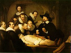
Rembrandt's "The Anatomy of Dr. Tulp."
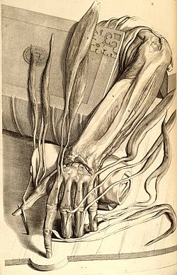
Gerard de Lairesse: Anatomical drawing of a left hand with tendons. From Govard Bidloo: Anatomy of the Human Body, Jacob van Poolsum, Utrecht, 1728 (reprint of the 1690 edition).
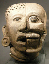
Mayan Bust
Workspaces
Macroscopic anatomy
Macroscopic anatomy deals with the structure of humans, animals or plants, namely with all things that can be seen with the naked eye. Here, not only externally visible structures are considered, but also in particular the structures that can be observed after cutting the body open and apart.
According to the type of approach macroscopic anatomy is divided:
- Descriptive anatomy is probably the most antiquated way of teaching anatomy. It teaches the individual structures of the body only in terms of their external appearance. Functional, topographical and systematic aspects are not considered. However, despite all its disadvantages, modern anatomy always has a descriptive component, because a doctor must be able to recognise pathological changes in an organ.
- Topographical anatomy describes the individual structures of the body according to their spatial positional relationships to each other (topos: Greek "place"). The great advantage is certainly that the doctor/veterinarian acquires a very application-oriented knowledge. For example, it is not only important for a hand surgeon to know to which larger organ system a structure belongs, he must know in particular where nerves, blood vessels or tendons run exactly. Topographic anatomical knowledge is also of great importance for the application of imaging techniques. Topographic anatomy uses standardized position and direction designations that are independent of the actual body position and instead uses relative reference points. Functional relationships can be deduced not only from structural and, in particular, topographical peculiarities of the structure of bodily form and organs, but also, and especially, from the somatotopic structure of nervous tissue.
- Systematic anatomy, which had replaced topographical anatomy in the 20th century, groups the individual structures of the body into functionally related organ systems. While this allows for some categorization and facilitates learning, it also has drawbacks. Topographical aspects, which the doctor/veterinarian has to deal with in everyday clinical practice, are not taken into account. In addition, all organ systems are also interconnected again, the skin has, for example, blood vessels, nerves, cells of the immune defence etc.
- Comparative anatomy studies the body structure of different animal species. Classical biological systematics already relied on structural similarities and differences for the classification from kingdoms to species, but increasingly genetic differences are also included in the classification. With the juxtaposition and comparison of different species, sometimes observations of a species can be interpreted in the first place. Furthermore, this comparison offers the possibility of recognizing certain basic structural principles and thus provides the basis for a common naming. The first lectures in the field of comparative anatomy were given by Johann Friedrich Blumenbach in 1785, and the physician, anatomist and physiologist Hermann Friedrich Stannius (1808-1883) from Rostock introduced the term zootomy, which almost entirely coincides with the term comparative anatomy.
Microscopic anatomy
Microscopic anatomy (histology) is responsible for the examination of anatomical structures below the level visible to the naked eye. It describes the fine structure of organs, tissues and cells.
Embryology
→ Main article: Embryology
Embryology describes the formation of anatomical structures during embryonic development. Based on the history of development, a variety of topographical and functional relationships can be identified. Embryological knowledge is also indispensable for understanding the development of malformations.
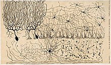
Microscopic section of the cerebellum of a chicken
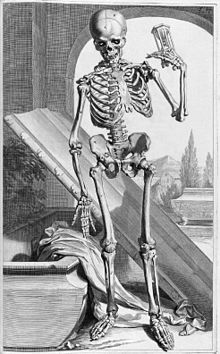
Illustration of the human skeleton by Gerard de Lairesse in Govard Bidloo's Ontleding des menschlyken lichaams of 1685. The vanitas symbols coffin and hourglass still justify the subject.
Questions and Answers
Q: What is anatomy?
A: Anatomy is the study of the structures and positions of organs, muscles, glands, and bones in the bodies of people and other animals.
Q: What does an anatomist do?
A: An anatomist is a person who studies anatomy.
Q: When did the study of human anatomy begin?
A: The study of human anatomy dates back to 1600 BC when Egyptians first began studying it.
Q: What did the Egyptians discover in their study of human anatomy?
A: The Egyptians discovered the functions of many organs, such as the liver, spleen, kidneys, heart, etc. They were the first to discover the structure and functions of the lymphatic system.
Q: Who usually employs academic human anatomists?
A: Academic human anatomists are usually employed by universities, medical schools, and teaching hospitals.
Q: What is gross anatomy?
A: Gross anatomy is the study of parts of the body that are big enough to see.
Q: What is micro-anatomy?
A: Micro-anatomy is the study of smaller parts of the body.
Search within the encyclopedia