Tissue (biology): structure, types, functions and study
A tissue is a group of similar cells that perform a specific role within multicellular organisms. Covers animal and plant tissues, organization, examples, histology and medical relevance.
A tissue is a collection of cells that share a common origin and collaborate to perform one or more specific functions within a multicellular organism. Tissues are the intermediate level of biological organization between cells and organs. Cells within a tissue often have similar form and specialized roles that together enable processes such as support, transport, protection, movement and information transfer.
Image gallery
6 Images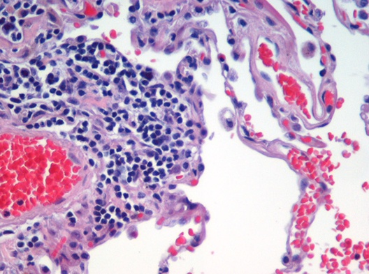


Characteristics and organization
Tissues are defined by their cellular composition, extracellular material and pattern of interaction. They may include a single cell type or several that work together. Boundaries between tissues are often visible under a microscope, and tissue architecture—arrangement of cells, fibers and fluid—determines mechanical and physiological properties. In animals, groups of different tissues combine to form an organ, such as the heart, which contains muscle, connective and epithelial components working in concert.
Major types in animals
- Epithelial tissue: sheets of cells that line surfaces and cavities, providing protection and selective transport.
- Connective tissue: diverse class including bone, cartilage, blood and adipose tissue; it provides support, binds structures and stores energy. See connective tissue.
- Muscle tissue: specialized for contraction and force generation; includes skeletal, cardiac and smooth types. See muscles.
- Nervous tissue: composed of neurons and supporting cells that transmit signals and process information. See nerves.
Plant tissues
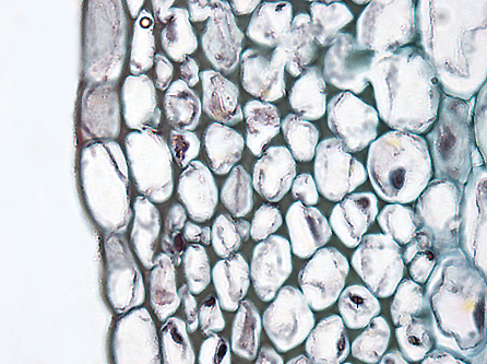
Plants organize tissues differently from animals. Broad categories include meristematic tissues (regions of active cell division) and permanent tissues: dermal (surface protection), ground (photosynthesis, storage and support) and vascular (transport of water, minerals and sugars via xylem and phloem). These systems enable growth, resource distribution and structural integrity in stems, roots and leaves.
Study and historical notes
The microscopic examination of tissues is called histology. Early observations of cells and tissues date to the 17th and 18th centuries; development of staining methods and more advanced optics expanded the field. Modern histology combines light and electron microscopy with molecular markers to reveal cellular detail and diagnose disease. Practical tools include simple stains, immunohistochemistry and digital imaging; samples are typically prepared and visualized with a microscope.
Functions, examples and significance
Tissues underpin nearly every organ system. For example, the cardiac muscle (myocardium) generates rhythmic contraction while internal connective linings (endocardium) and external membranes (pericardium) support and protect the heart. Valves and associated structures guide blood flow through coordinated tissue action (valves). Understanding tissue structure is vital in medicine for recognizing injury, inflammation, degeneration and cancer, and it is central to regenerative medicine and tissue engineering research.
Distinctions and notable facts
- Tissues differ in regenerative capacity: some, like liver or epithelial linings, repair well, whereas cartilage and certain neurons regenerate poorly.
- Pathology often appears first at the tissue level, making histological examination essential for diagnosis.
- Tissue classification is a practical framework; at the molecular level, cell types show continuous variation and plasticity.
For further introductory reading on organs and tissue function, follow resources on general anatomy and histology, or consult specialist texts linked through academic and medical portals: definitions and origins and broader references on muscle and nerve structure are commonly available.
Related articles
Author
AlegsaOnline.com Tissue (biology): structure, types, functions and study Leandro Alegsa
URL: https://en.alegsaonline.com/art/100102