
Tongue: anatomy, functions, sensation and clinical overview
Concise encyclopedia entry on the human tongue: structure, surface features, taste, roles in eating and speech, innervation, blood supply, common conditions and basic clinical and hygiene notes.
Overview
The tongue is a muscular organ that occupies most of the oral cavity and contributes to taste, food manipulation, swallowing and speech. It is covered by a moist mucous membrane and includes both intrinsic muscles, which alter its shape, and extrinsic muscles, which change its position. For general anatomical context see anatomy resources and the relation to the rest of the mouth at oral cavity overview.
Image gallery
10 Images


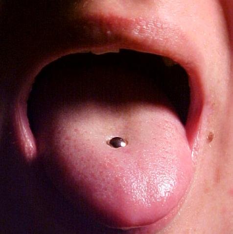



Surface, papillae and taste
The dorsal surface of the tongue is characterized by papillae of several types. Filiform papillae are numerous and provide friction but do not contain taste buds. Fungiform, foliate and circumvallate papillae contain taste receptors commonly called taste buds. Taste perception arises from chemoreceptors that detect basic qualities such as sweet, salty, sour, bitter and umami; the older idea of a strict "taste map" on the tongue is inaccurate, because taste receptors are distributed across the surface. For introductory material on taste and sensation see taste and sensation and more on receptor distribution at taste bud information.
Anatomy and supporting tissues
Intrinsic muscles (superior and inferior longitudinal, transverse and vertical) change the tongue's shape. Major extrinsic muscles include the genioglossus, hyoglossus, styloglossus and palatoglossus, which anchor the tongue to surrounding bones and soft tissues. The mucosa over the base of the tongue includes lymphoid tissue (lingual tonsil) that contributes to immune defense.
Innervation and blood supply
Sensory and motor innervation involve several cranial nerves. General sensation from the anterior two-thirds is carried by the lingual nerve (branch of the mandibular nerve), while taste from that region is carried by the chorda tympani (a branch of the facial nerve). The posterior third receives both taste and general sensation via the glossopharyngeal nerve. Motor control of most tongue muscles is by the hypoglossal nerve, with the palatoglossus muscle supplied by the vagus nerve. The main arterial supply is from branches of the lingual artery; venous drainage follows lingual veins toward the internal jugular system.
Functions
- Taste: Chemical sensing that informs food choice and safety.
- Mastication and bolus formation: Positioning, mixing with saliva and shaping of food for swallowing.
- Deglutition (swallowing): Propulsion of the bolus into the pharynx under coordinated control with the pharyngeal muscles.
- Speech and articulation: Precise tongue movements form many consonants and vowels; see further reading on articulation at speech production.
Development, variation and comparative notes
The tongue develops from several pharyngeal arches, which explains its complex innervation. Its size, shape and surface pattern vary between individuals and species. In animals, tongues may be adapted for lapping, grasping, or specialized feeding behaviors.
Clinical significance and care
Common conditions include inflammation (glossitis), fungal infection (oral candidiasis), geographic tongue (benign migratory glossitis), ankyloglossia ("tongue-tie") and neoplasms such as squamous cell carcinoma. Macroglossia (enlarged tongue) can affect breathing and speech and may contribute to sleep-disordered breathing. Examination of the tongue is a routine part of oral and general physical assessment: clinicians inspect color, texture, lesions, mobility and strength. Persistent pain, ulcers that do not heal, unexplained lumps or sudden changes in taste or mobility warrant medical evaluation. Basic care includes good oral hygiene, hydration and avoidance of irritants; specialist interventions range from conservative therapy to surgical procedures such as frenotomy or partial glossectomy in selected cases.
Notable points
Assertions that the tongue is the "strongest" muscle depend on the measure used and are not precise. The organ performs multiple coordinated tasks that make it essential for nutrition, communication and taste. For further trusted clinical summaries and images consult professional anatomy and oral health sources such as anatomy resources and oral health pages.


Structure
The tongue is divided into the tip (apex linguae), the body (corpus linguae) and the root (radix linguae). The upper surface of the tongue (the dorsum of the tongue, dorsum linguae) is somewhat convex, lies entirely exposed, and shows at the back a triangular depression, the blind hole, in which several mucous glands open. In phonetics, the anterior, mobile part of the dorsum of the tongue directly behind the tip of the tongue, which is involved in sound formation, is called the tongue blade. In the area of the tip of the tongue, a small salivary gland, the lingual tip gland (glandula lingualis anterior, Blandin-Nuhn gland), is located under the mucous membrane. In the rear area of the tongue, further salivary glands, the lingual glands (glandulae linguales), are located.
The lower surface is attached to the floor of the oral cavity with its central part and is attached at the front by a fold of the oral mucosa, the lingual frenulum (frenulum linguae), in such a way that only the tip of the tongue (apex linguae) and the lateral edges are free. If the lingual frenulum extends too far forward, the free movement of the tongue is impaired (ankyloglosson). This can impair sucking and sound production. This developmental disorder can be eliminated by an incision in the lingual frenulum (tongue loosening).
The rearmost, thickest part of the tongue, the root of the tongue (radix linguae), is attached to the hyoid bone, which lies in the upper part of the neck and is connected to the larynx again by muscles and ligaments.
In the midline of the tongue is a kind of vertical septum, the septum linguae. In all other respects, however, the tongue consists mainly of muscle fibres with numerous nerves and blood vessels running between them. The muscle fibres are arranged in all three spatial directions and thus enable the extremely high mobility of the tongue.
Tongue muscles
→ Main article: Tongue muscles
The tongue musculature is divided morphologically but also functionally-motorically into an inner tongue musculature - it runs entirely inside the tongue body - and an outer tongue musculature. The latter connects the tongue with the surrounding organ and tissue structures.
The internal muscles of the tongue (abbreviation M. corresponds to musculus) include the:
Shortening and widening of the tongue, lifting the tip of the tongue:
- superior longitudinalis
- inferior longitudinal muscle
Lengthening or narrowing of the tongue, sticking out the tip of the tongue:
- transverse lingual muscle
- lingual verticalis
and the external muscles of the tongue include:
Pull the tongue down in front (ventro-caudal):
- M. genioglossus
Pull the tongue down behind (dorso-caudal):
- M. chondroglossus
Pull the tongue up backwards (dorso-rostral):
- M. styloglossus
Pull the tongue down behind (dorso-caudal):
- M. hyoglossus
Narrowing of the isthmus faucium:
- palatoglossus


Innervation
The tongue is innervated in a very complex way by several nerves:
- The tongue is supplied motorically by the hypoglossal nerve.
- Sensory and sensory innervation occurs in the posterior third of the tongue through the glossopharyngeal nerve.
- The sensory supply of the anterior two-thirds is provided by the lingual nerve.
- The taste stimuli of the anterior two-thirds of the tongue are transmitted by the chorda tympani, a branch of the facial nerve.
Related articles
Author
AlegsaOnline.com Tongue: anatomy, functions, sensation and clinical overview Leandro Alegsa
URL: https://en.alegsaonline.com/art/100530
Sources
- strange-facts.info : interesting tongue fact
- commons.wikimedia.org : Tongues