
Histology: The Microscopic Study of Cells and Tissues
Histology examines the microscopic structure of plant and animal tissues. This article outlines methods, history, applications in research and medicine, and how histology differs from related fields.
Histology is the branch of biological science that examines the fine structure of cells and tissues as seen with microscopes. Often described as microscopic anatomy, it includes the study of tissues from both plants and animals. Histology overlaps with but is distinct from cytology, which focuses mainly on individual cells. It is a foundational tool in modern biology and medicine, where tissue structure informs function, development, and disease.
Image gallery
10 Images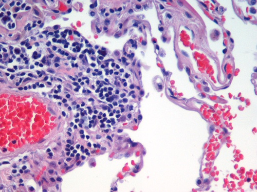




Principles and common methods
Preparing tissue for microscopic study requires several standard steps: fixation to preserve structure, embedding to provide support, thin sectioning, and staining to highlight components. Stains reveal different chemical or structural features; for example, routine dyes differentiate nuclei and cytoplasm, while specialized labels detect proteins or carbohydrates. Observations are made with light-based instruments or higher-resolution devices depending on scale.
- Optical techniques: Conventional observation typically uses a light microscope, including brightfield, phase contrast, and fluorescence variants.
- Electron microscopy: For ultrastructural detail, the electron microscope resolves organelles and macromolecular arrangements beyond the light microscope's limits.
- Modern extensions: Immunohistochemistry and in situ hybridization combine molecular probes with histology to map proteins and nucleic acids in their tissue context.
History and development
Histology grew in the 19th century as lenses and staining techniques improved, enabling scientists to move beyond gross anatomy and describe tissue organization systematically. Over time, methodological advances—paraffin embedding, microtomy, fluorescent tags, and electron optics—transformed it from descriptive work into a quantitative discipline integral to cell biology and pathology.
Applications and importance
Histology is used across research and clinical practice. In research, it documents normal development, tissue remodeling, and responses to experimental treatments. Clinically, histological examination of biopsies and surgical specimens is central to diagnosing cancers, inflammatory diseases, and many other conditions (a subspecialty called histopathology). Educational programs use histology to teach how microscopic structure relates to organ function.
Distinctions and notable points
Key distinctions include the difference between cytology and histology (cells versus organized tissue architecture) and between routine histology and histopathology (diagnostic emphasis). Modern histology often integrates imaging, molecular labeling, and digital analysis to quantify patterns that were once assessed qualitatively. The field continues to evolve as new markers, imaging modalities, and computational tools expand what can be observed and measured.
For further general reference on terminology and methods, see introductory resources on microscopic anatomy, comparisons with cytology, or applied perspectives in biology and medicine. Practical technique guides often discuss light and electron microscopy in detail (light microscope, electron microscope).
Questions and answers
Q: What is histology?
A: Histology is the study of the microscopic anatomy of cells and tissues of plants and animals.
Q: What is the focus of histology?
A: The focus of histology is on the tissues, although it is also a part of cytology.
Q: Why is histology considered an essential tool in biology and medicine?
A: Histology is considered an essential tool in biology and medicine because it allows researchers and medical professionals to understand the structure and function of tissues at the microscopic level.
Q: What is the usual method of conducting histological studies?
A: The usual method of conducting histological studies is by looking at cells and tissues under a light or electron microscope.
Q: What is the importance of the preparation of the tissue in histology?
A: The preparation of the tissue is important in histology because it allows researchers to obtain a clear and accurate image of the tissue under the microscope.
Q: What is the role of light microscope and electron microscope in histology?
A: The role of light microscope and electron microscope in histology is to magnify the tissues and cells being studied to make them easier to visualize and analyze.
Q: How has histology contributed to the understanding of biology and medicine?
A: Histology has contributed significantly to the understanding of biology and medicine by allowing researchers and medical professionals to study the structure and function of tissues at the microscopic level, including the diagnosis and treatment of diseases.
Related articles
Author
AlegsaOnline.com Histology: The Microscopic Study of Cells and Tissues Leandro Alegsa
URL: https://en.alegsaonline.com/art/44434