
Microscope: principles, types, history and common uses
Comprehensive overview of microscopes: how they magnify, main designs and components, historical development, typical applications in science and medicine, and practical limitations and care.
A microscope is a precision instrument designed to magnify objects that are too small to see clearly with the unaided eye. By collecting and redirecting light (or other kinds of radiation), microscopes create an enlarged image that reveals shape, structure and detail. They are indispensable tools in biology, medicine, materials science and education, and they range from simple single-lens devices to elaborate systems combining optics, electronics and computation. For an introduction to practical use and safety, beginners often consult educational resources such as user guides.
Image gallery
10 Images





Basic components and how they work
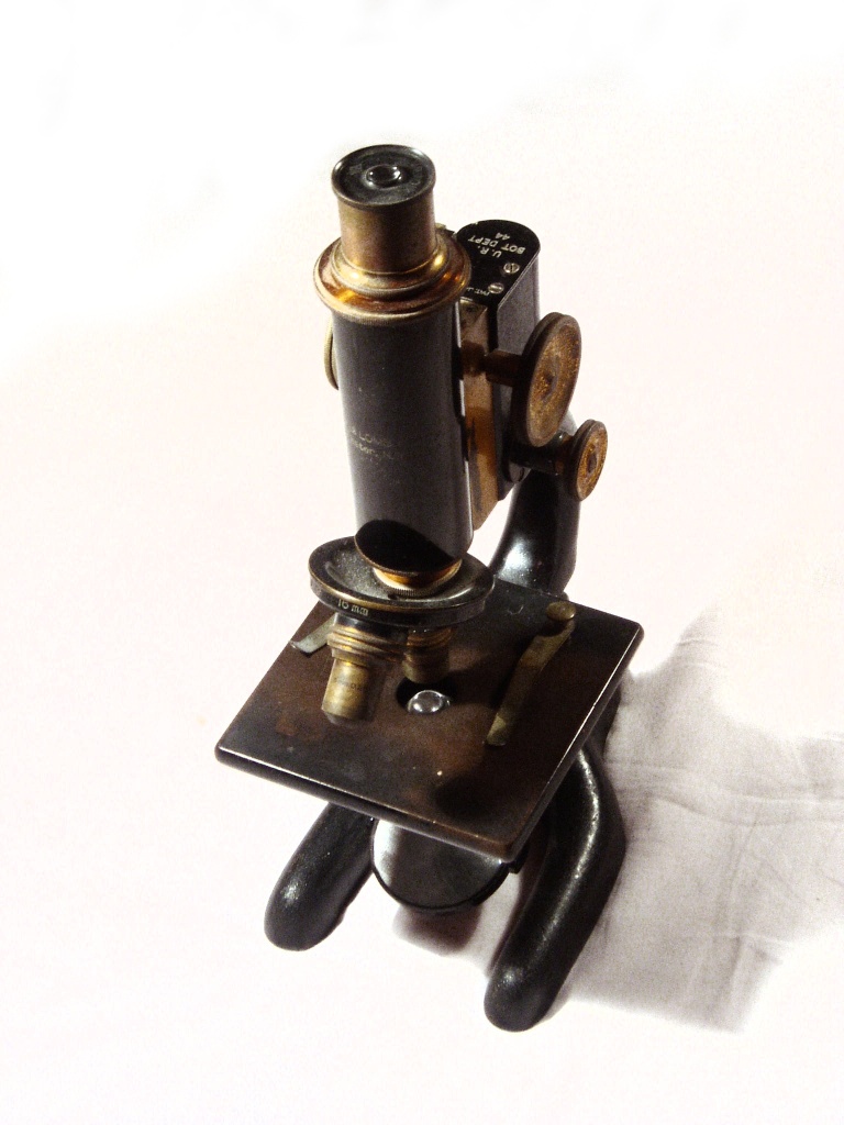
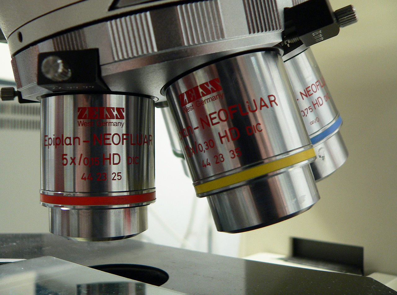
Most optical microscopes share a few common parts: an illumination source, a stage to hold the specimen, objective lenses close to the sample, and an eyepiece or camera to view the image. In compound microscopes the image is formed in two steps: the objective produces a magnified intermediate image, and the eyepiece enlarges that image for the observer. Total magnification is the product of the objective and eyepiece magnifications. Resolution — the ability to distinguish two nearby points — depends on the lens quality and the wavelength of light used, and it ultimately limits how much detail can be resolved.
Major types of microscopes
- Simple microscopes: use a single lens or element, similar to a magnifying glass; useful for quick inspection and historical instruments.
- Compound light microscopes: common in classrooms and labs; provide high magnification with multiple objective lenses and transmitted light.
- Stereomicroscopes (dissecting scopes): offer lower magnification but a three-dimensional view for dissection or inspection of larger specimens.
- Electron microscopes: replace light with electron beams to reach much higher resolution for observing ultrastructure; they require vacuum and special sample preparation.
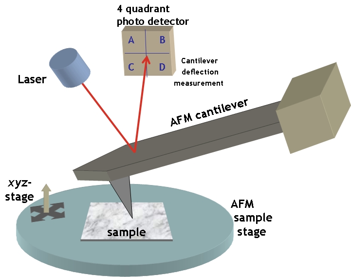
- Scanning probe and other advanced microscopes: use physical probes or non‑optical signals to map surfaces at the nanoscale.
History and development
Early magnifying lenses were used for reading and simple observation; the first microscopes that could be called scientific instruments appeared in the late 16th and early 17th centuries. Over the following centuries improvements in lens grinding, illumination, and mechanical design led to compound microscopes capable of routine laboratory use. In the 20th century the introduction of electron optics and, later, confocal and digital imaging expanded the range of structures that could be examined and the ways images are recorded and analyzed.
Applications and examples
Microscopes underpin many scientific and clinical tasks. In biology they reveal cells, organelles and microorganisms; in medicine they assist diagnosis through examination of tissue sections or blood smears; in materials science they expose grain structure, defects and coatings. Educational microscopes are widely used in classrooms to introduce students to cellular life and basic microscopy techniques. Professional workflows often pair microscopes with cameras and software for measurement, documentation and remote collaboration — resources that can be found in teaching materials and supplier catalogs such as laboratory pages and academic collections.
Care, limitations and notable distinctions
Proper handling and maintenance — cleaning lenses, avoiding knock or contamination, and using appropriate mounting media — preserve optical performance. Optical microscopes are limited by light wavelength: beyond a certain magnification, images become larger but not more detailed. Electron and scanning probe microscopes overcome optical limits but introduce higher cost, more complex sample preparation and operational constraints. For practical comparisons and purchasing decisions, many users consult reviews and buying guides available from manufacturers and educational institutions, for example comparison guides and technical notes.
Whether used to discover new microbes, to inspect integrated circuits or to teach basic scientific observation, the microscope remains a foundational instrument. For further introductory demonstrations, tutorials and troubleshooting steps, readers can follow instructional resources such as online tutorials.
Questions and answers
Q: What is a microscope?
A: A microscope is a scientific instrument that makes small objects look larger, allowing people to see small things.
Q: Who typically uses microscopes in their jobs?
A: Doctors and scientists are among the people who frequently use microscopes in their jobs.
Q: What kinds of science students use microscopes in their studies?
A: Students in science classes such as biology also use microscopes to study small things.
Q: What is the difference between a simple microscope and a compound microscope?
A: A simple microscope has only one lens, while a compound microscope has at least two lenses.
Q: What are the names of the lenses in a compound microscope?
A: The lens closer to the eye is called the eyepiece, while the lens at the other end is called the objective.
Q: How do the lenses in a compound microscope work together to magnify objects?
A: The lenses multiply up, so a 10x eyepiece and a 40x objective together give 400x magnification.
Q: Compared to a magnifying glass, how strong can a microscope magnify objects?
A: Microscopes can make things seem larger than they are, to about 1000 times larger, which is much stronger than a magnifying glass that works as a simple microscope.
Related articles
Author
AlegsaOnline.com Microscope: principles, types, history and common uses Leandro Alegsa
URL: https://en.alegsaonline.com/art/64613
Sources
- apnews.excite.com : "2 Americans, 1 German win chemistry Nobel"
- nytimes.com : "2 Americans and a German Are Awarded Nobel Prize in Chemistry"