
Tibia (shinbone): anatomy, function, development and clinical significance
The tibia, or shinbone, is the larger weight-bearing bone of the lower leg. This article explains its anatomy, growth, functions, common injuries and how it differs from the fibula.
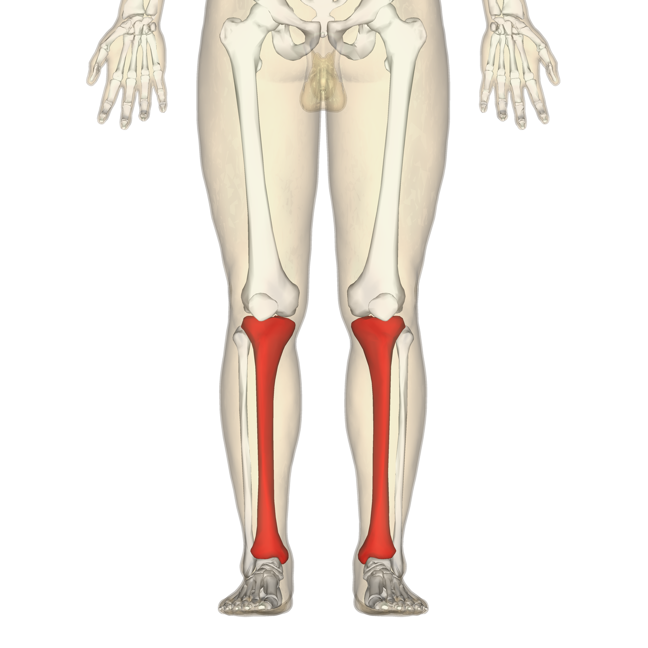
The tibia, commonly called the shinbone or shankbone, is the larger and stronger of the two long bones in the lower leg. Situated below the knee and running down to the ankle, the tibia lies parallel to the fibula and transmits most of the load from the upper limb of the body onto the foot. It is present in many vertebrates and is a key element of upright posture and terrestrial locomotion.
Image gallery
10 Images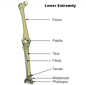


Anatomical features
The tibia has a triangular cross-section and three borders. Its main parts are the proximal end (with medial and lateral condyles and the tibial plateau), the shaft, and the distal end (which includes the medial malleolus). Surface landmarks and attachments include the tibial tuberosity, for patellar ligament attachment, and numerous muscular and interosseous membrane attachments along the shaft. Key functional regions:
- Proximal tibia: weight-bearing articular surface that forms part of the knee joint.
- Shaft: stiff, load-bearing column with muscular attachments and nutrient foramen.
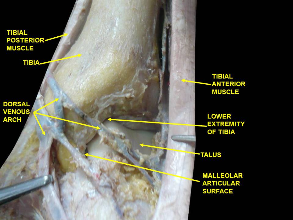
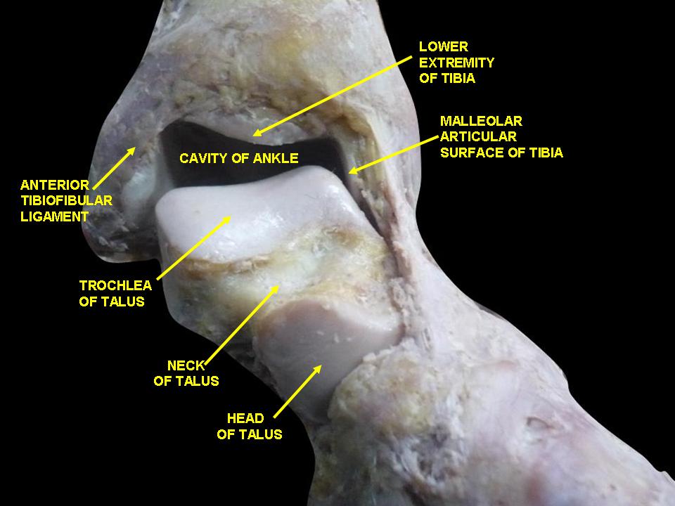
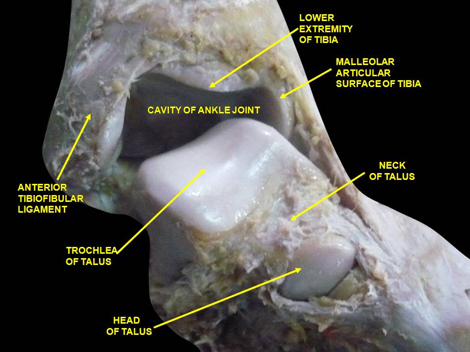
- Distal tibia: contributes to the ankle joint and provides the medial malleolus.
Development and variation
The tibia develops from cartilage in the fetus and ossifies through primary and secondary growth centers during childhood and adolescence. Growth plates (physes) at the ends allow lengthening; injuries to these plates can alter final bone length. Size and shape vary by age, sex and species; in humans the tibia is the second largest bone of the body and is adapted for bearing a large share of body weight.
Function and biomechanics
The tibia’s primary role is load transmission and support during standing, walking, running and jumping. It forms the major articular surfaces of the knee and contributes to the ankle mortise, stabilizing the lower limb. Muscles of the anterior, lateral and posterior compartments attach to the tibia and act to move the foot and knee, coordinate balance, and absorb impact during gait. The tibia works together with the fibula and soft tissues to maintain limb alignment and joint congruity.
Clinical relevance
Tibial fractures are common and range from stress fractures in athletes to high-energy fractures that may involve the joint surface. Typical issues include tibial shaft fractures, tibial plateau fractures, and distal fractures involving the medial malleolus. Treatment varies from conservative casting to surgical fixation. Growth-plate injuries, osteomyelitis, and compartment syndrome are additional concerns. Anatomical knowledge of the tibia is essential for imaging, orthopedic surgery and rehabilitation.
Comparisons, evolution and notable facts
Compared with the fibula, the tibia bears most of the axial load and forms major joints; the fibula contributes to lateral stability and muscle attachment. Across vertebrates, the tibia shows adaptations to different gaits and habitats. The tibia is fundamental to human mobility and is involved in everyday movement such as standing, walking and running. For additional anatomical reference and clinical images see bone atlas and standard orthopedic texts on the lower limb.
Related articles
Author
AlegsaOnline.com Tibia (shinbone): anatomy, function, development and clinical significance Leandro Alegsa
URL: https://en.alegsaonline.com/art/99778