Stethoscope
A stethoscope is an acoustic or electronic instrument used in auscultation to listen to internal body sounds—heart, lungs, bowel and blood flow—by clinicians, emergency responders and veterinarians.
The stethoscope is a diagnostic instrument used to listen to sounds produced within the body, a practice called auscultation. It allows clinicians and other caregivers to detect or monitor heartbeats, breath sounds, bowel noises and vascular murmurs without invasive procedures. Because it is portable, inexpensive compared with imaging equipment, and easy to use with training, the stethoscope remains a central tool in physical examination and bedside assessment. For more background on basic designs and uses see stethoscope overview.
Image gallery
10 Images


Parts and common variants
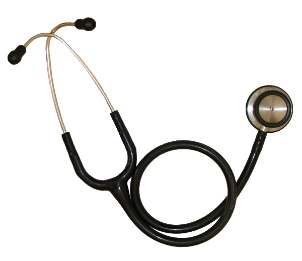
Modern stethoscopes typically have several distinct components: a chestpiece (with a diaphragm and often a bell), flexible tubing, binaural metal tubes, and earpieces. The diaphragm is a flat, membrane-covered surface that transmits higher-frequency sounds; the bell is shaped like a cup and is better for low-frequency sounds. Tubing carries acoustic vibrations from the chestpiece to the listener’s ears; binaurals allow adjustment of angle and fit.
- Acoustic stethoscopes: traditional models that transmit sound mechanically.
- Electronic stethoscopes: amplify sound and may offer filtering, recording, or Bluetooth output.
- Specialized forms: pediatric, neonatal, cardiology variants with different chestpiece sizes or enhanced sensitivity, and simple obstetric/fetal devices.
These instruments are used by physicians, nurses, paramedics, and veterinarians; various professions may prefer specific models or accessories depending on clinical needs and patient size. See resources for professional guidance at clinical users.
How it is used and what it can reveal
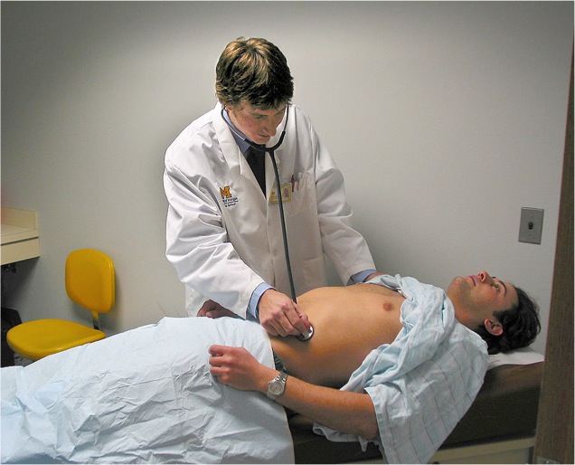
Auscultation technique involves placing the chestpiece on defined anatomical sites while the patient breathes normally or performs maneuvers. Typical listening areas include the chest over heart valves, the anterior and posterior lungs, and the abdomen for bowel sounds. Clinicians listen for characteristics such as rhythm, intensity, pitch and timing. Examples of diagnostically useful findings are heart murmurs suggesting valve disease, crackles that may indicate fluid in the lungs, wheezes associated with airway narrowing, and bruits from turbulent blood flow in arteries.
Stethoscopes also assist in measuring blood pressure (listening for Korotkoff sounds when using a sphygmomanometer) and in bedside triage. Electronic models can record and share sounds for consultation or teaching; however, interpretation depends heavily on practitioner skill and clinical context. For practical guidance on technique and interpretation, consult auscultation resources.
History and development
The stethoscope was invented in 1816 by the French physician René Laënnec, who initially used a rolled paper tube to listen to the chest and later built a wooden monaural instrument. His work formalized auscultation and linked clinical sounds with pathological findings at autopsy. Over the nineteenth and twentieth centuries the device evolved into binaural metal and rubber tubing designs and then to modern materials and electronic amplification. Historical context and biographies are available at historical references.
Limitations, infection control and cultural role
While valuable, the stethoscope has limits: ambient noise, improper technique, and subtle pathology can reduce diagnostic accuracy. It is an adjunct to, not a replacement for, imaging and other tests. Infection control practices—cleaning diaphragms and earpieces between patients and avoiding contact with open wounds—are important to reduce cross-contamination. Many institutions publish cleaning protocols and recommendations; see institutional guidance at stethoscope hygiene.
Beyond clinical function, the stethoscope has symbolic value as an emblem of healthcare professionals and remains a core element of medical education and physical diagnosis training. Its design continues to adapt with materials science and digital technology, but the fundamental purpose—listening to the living body—remains unchanged.
Related articles
Author
AlegsaOnline.com Stethoscope Leandro Alegsa
URL: https://en.alegsaonline.com/art/93818