
Rectum (anatomy, function, and clinical importance)
Overview of the rectum: structure, role in defecation, temperature measurement, common conditions, diagnostic procedures, and key anatomical relationships.
The rectum is the final straight segment of the large intestine and a central part of lower gastrointestinal anatomy. It connects the sigmoid colon to the anus and acts as a temporary reservoir for fecal material before elimination. The name derives from Latin and reflects the organ's relatively direct course toward the body's exit.
Image gallery
10 Images


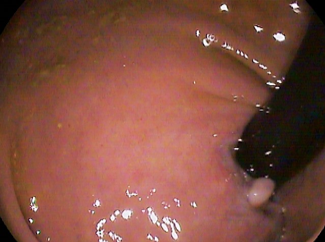

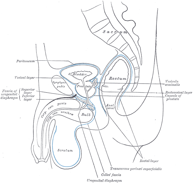
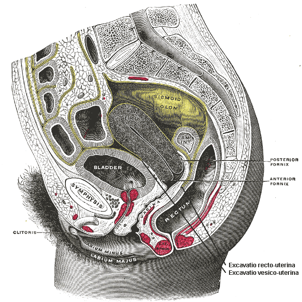

Anatomical features
In adults the rectum is approximately 10–15 cm long and lies within the pelvis. Its upper portion is continuous with the sigmoid colon, and its lower end narrows into the anal canal. The rectal ampulla, an expanded region near the end, accommodates stool before defecation. Wall layers include mucosa, submucosa, muscular layers and serosa/adventitia similar to other large bowel segments. Two sphincters around the anal canal — the internal sphincter (smooth muscle) and the external sphincter (skeletal muscle under voluntary control) — regulate continence.
Physiology and function
The rectum stores feces and triggers the defecation reflex when distended. Sensory receptors in the rectal wall distinguish solid, liquid or gaseous contents and help coordinate voluntary relaxation of the external sphincter with pelvic floor muscles. The rectum also provides access for certain medical measurements; for example, body temperature can be measured rectally, which typically yields readings slightly higher than those taken orally in the mouth.
Blood supply, innervation and clinical relevance
Arterial supply arises from branches of the inferior mesenteric and internal iliac systems; venous drainage has connections to both the portal and systemic circulations, a clinically relevant arrangement for conditions such as hemorrhoids. Innervation includes autonomic fibers that mediate reflexes and somatic fibers (via the pudendal nerve) that control voluntary sphincter contraction and sensation.
Common conditions and clinical assessment
- Disorders: hemorrhoids, anal fissures, inflammatory conditions (proctitis), rectal prolapse and malignancies affecting the rectum are among frequent clinical concerns.
- Screening and diagnosis: evaluation may use digital rectal examination, anoscopy/proctoscopy and endoscopic techniques such as sigmoidoscopy or colonoscopy. Biopsy and imaging support diagnosis when structural disease is suspected.
- Management: ranges from conservative measures and topical treatments to surgical procedures, depending on the condition and severity.
Development, history and distinctions
Embryologically, the rectum develops from the terminal portion of the hindgut. Historically and anatomically it is distinguished from the sigmoid colon by its fixed pelvic position and from the anal canal by transition of epithelial types and sphincter anatomy. Specialists who focus on this region include colorectal surgeons and gastroenterologists; primary-care clinicians commonly perform screening and basic examinations.
For further general context about adjacent structures and related topics, see resources on the large intestine, the anus, pelvic anatomy (pelvic structures), and clinical guidance from recognized sources such as professional societies or review articles (historical and clinical references). Practical information and patient guidance often link rectal function to bowel habits, continence, and when to seek medical evaluation.
Related articles
Author
AlegsaOnline.com Rectum (anatomy, function, and clinical importance) Leandro Alegsa
URL: https://en.alegsaonline.com/art/81610