Paranasal sinuses: structure, function, development, and clinical relevance
Paranasal sinuses are air-filled cavities in facial bones that communicate with the nasal cavity; they are lined by mucosa and have roles in respiration, voice resonance, skull lightening, and disease.
The paranasal sinuses are air-filled spaces located within the bones around the nose that open into the nasal cavity. They are generally considered part of the respiratory system because they are lined by a mucous membrane and contribute to conditioning inhaled air. The lining is a specialized epithelium that produces mucus and moves trapped particles toward the nasal passages. These cavities develop by pneumatization of bone, a process that begins in childhood and continues into adolescence.
Image gallery
7 Images


Anatomy and principal sinuses
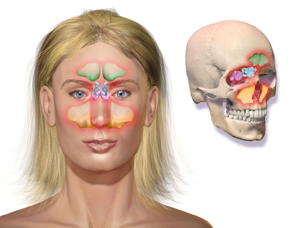
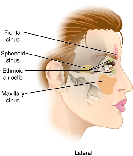
Four paired sinuses are conventionally recognized in human anatomy: the maxillary, frontal, ethmoid and sphenoid sinuses. Each occupies a discrete region of the facial and cranial bones and drains into specific areas of the nasal cavity. The sinuses are lined by a mucous membrane that is a type of mucous membrane and lies against the surrounding bones. Their internal surface is ciliated and produces mucus that helps trap particles and pathogens.
Proposed functions and biological roles
Because air-filled sinuses occur in many land vertebrates, several hypotheses have been proposed for their functions. Likely contributions include:
- Lightening the skull by creating hollow spaces within otherwise solid bones, which reduces mass without greatly increasing volume.
- Humidifying and warming inhaled air via the mucosal surface, aided by epithelial cilia and mucus transport.
- Modifying voice resonance and contributing to the characteristic timbre of an individual's voice.
- Serving as a first-line immunological surface that traps particles and contains immune cells in the mucosa.
Evolutionary and comparative context
Air-filled cranial and facial cavities are found across a wide range of vertebrates. Examples appear among mammals, many birds, and groups of reptiles; fossil evidence shows similar pneumatic features in some dinosaurs and in living crocodilians. The distribution and complexity of these spaces vary by lineage and are interpreted as adaptations that balance structural strength, metabolic cost, and respiratory or sensory demands.
Development and variation
Pneumatization starts as outpouchings of the nasal mucosa into adjacent bone. The timing and extent differ between individuals; some people have extensive pneumatization with additional accessory cells, while others have small or asymmetric sinuses. Anatomical variations influence drainage patterns and susceptibility to obstruction.
Clinical relevance and common disorders
Because the sinuses communicate with the nasal cavity through narrow openings, they are prone to blockage and infection (sinusitis). Chronic inflammation can cause pain, reduced sense of smell, and mucus retention; complications include mucoceles and spread of infection to nearby structures. The maxillary sinus is noteworthy for its proximity to upper teeth, so dental infections can affect it. Treatments range from medical therapy to endoscopic surgical procedures that restore drainage and ventilation.
Distinctions and notable facts
Although often discussed together, the paranasal sinuses are distinct from the central nasal passages; they are separate cavities that communicate via ostia. Their exact contributions to human physiology are still debated, and multiple functions may act together rather than a single dominant role. For further general reading on related anatomy and physiology, see general resources on the nasal cavity and the respiratory system, as well as specialized sources on mucosal biology and clinical management techniques such as endoscopic sinus surgery.
Reference links and introductory materials can be consulted for deeper study: mucous membrane overview, cranial bone anatomy, mammalian respiratory structures, avian air sac and sinus relationships, pneumatic bones in fossils, crocodilian skull pneumatization, epithelial cell types, and mucus biology.
Related articles
Author
AlegsaOnline.com Paranasal sinuses: structure, function, development, and clinical relevance Leandro Alegsa
URL: https://en.alegsaonline.com/art/74566