Endoskeleton: internal support systems in animals
An endoskeleton is an internal structural system that supports an animal's body. This article explains types, development, examples, advantages, and how endoskeletons differ from exoskeletons.
Overview
An endoskeleton is an internal framework of structural elements that supports an animal's body, anchors muscles and protects some internal organs. Unlike external armour, an endoskeleton occupies space within tissues. The term is used for a wide range of internal support strategies found across the animal kingdom, from the mineralized bones of vertebrates to the calcareous plates of echinoderms and the microscopic spicules of some sponges. For a general term, see animals and how they build internal support.
Image gallery
9 Images


Basic characteristics and components
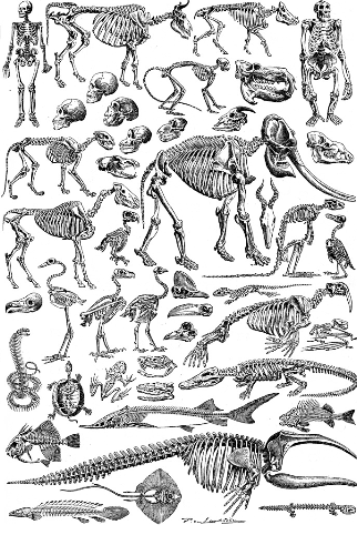
Endoskeletons can be built from different materials and organized in distinct ways. In vertebrates the endoskeleton is primarily composed of bone and cartilage. Cartilage is a flexible connective tissue often present in early development and in adult structures (for example, the shark skeleton). Bone is a mineralized tissue that provides rigid support and can remodel throughout life. Typical components include:
- Bones — rigid, mineralized elements with marrow and blood supply.
- Cartilage — flexible support found in joints, ears, and some fish.
- Ligaments and tendons — connective tissues that stabilize joints and transmit muscle forces.
- Joints and growth plates — enable movement and postnatal size increase.
Other forms of internal support
Not all internal supports are homologous to vertebrate bone. Echinoderms, for example, possess a true internal skeleton formed from calcareous ossicles beneath the skin; see Echinodermata. Some sponges (phylum Porifera) rely on a mesh of spicules and spongin fibers which function as an internal scaffold. A different situation occurs in some cephalopods: members of the subclass Coleoidea show retention or internalization of ancestral shell material rather than a mesoderm-derived skeleton.
Development and evolutionary notes
A true endoskeleton, in the strict developmental sense, is derived from the mesoderm germ layer. This applies to vertebrates and echinoderms where skeletogenesis involves mesodermal cells that secrete mineralized matrix. In contrast, internalized shell elements in some molluscs are modifications of an exoskeletal structure. The molluscan lineage that gave rise to internalized shells did not evolve a mesodermal skeleton but rather developed internal structures by transforming an ancestral outer shell. The distinction between a mesodermal skeleton and an internalized exoskeleton is important for evolutionary interpretation; see examples such as the internalized 'cuttlebone' in cuttlefish and other cephalopods.
Functions, advantages and limitations
Endoskeletons provide structural support, permit larger body sizes, and offer internal attachment surfaces for muscles that produce efficient leverage for movement. They also allow continuous growth without the need for periodic shedding, a key advantage over a rigid external shell (exoskeleton). However, endoskeletons generally offer less direct protection from external injury or predators than thick armor, so many animals combine skeletal support with behavioural, physiological, or integumentary defenses.
Examples and notable distinctions
- Vertebrates: humans and other mammals have an ossified skeleton with marrow-filled bones; sharks have predominantly cartilaginous skeletons.
- Echinoderms: sea stars and sea urchins possess internal calcareous ossicles forming a test or endoskeleton.
- Poriferans: sponge spicules form a micro-scale internal lattice used for support.
- Coleoid cephalopods: some species possess internalized shell elements such as the cuttlebone; these arose from an ancestral mollusc shell rather than from mesoderm, so some authors distinguish these structures from a true skeleton.
For further reading and comparative anatomy resources, consult general texts and specialist articles: use the following as starting references: animals overview, taxonomy pages at phyla, or focused entries on Coleoidea and echinoderms (Echinodermata).
Related articles
Author
AlegsaOnline.com Endoskeleton: internal support systems in animals Leandro Alegsa
URL: https://en.alegsaonline.com/art/31396