
Right upper quadrant (abdomen)
Anatomical and clinical overview of the right upper quadrant (RUQ) of the abdomen: boundaries, typical organs, examination clues, common causes of RUQ pain and important distinctions for diagnosis.
Overview
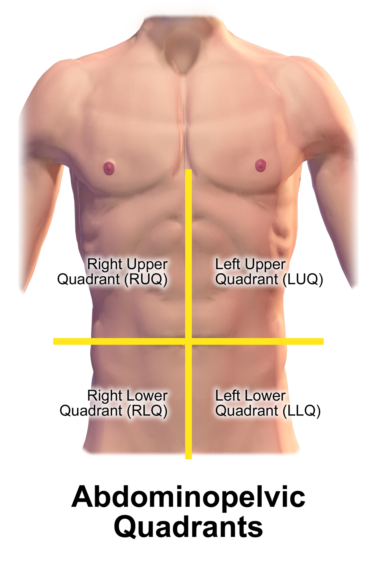
The right upper quadrant (RUQ) is one of four standard divisions used in clinical abdominal assessment. It is defined by two imaginary lines: a vertical midline (median plane) and a transverse line that passes through the umbilicus. The RUQ occupies the upper-right portion of the abdomen and is a practical, simple way for clinicians to communicate where discomfort, masses, or other findings are located. It should not be equated with the epigastrium, which is the central upper region between the right and left upper quadrants.
Image gallery
6 Images



Boundaries and anatomical contents
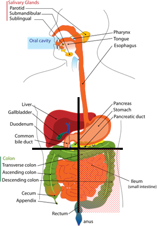
Surface boundaries are practical rather than precise: superiorly the lower margin of the thorax (right rib cage), medially the median plane, and inferiorly roughly at the level of the umbilicus. Because organs occupy three-dimensional space and shift with posture, respiration, body habitus and pregnancy, the organs described below may extend beyond the RUQ in some people.
- Liver (mainly the right lobe) — often the largest organ felt in this area.
- Gallbladder — tucked under the liver and a frequent source of RUQ pain.
- Duodenum (first and second parts) — part of the small intestine that lies near the liver and pancreas.
- Pancreas (head) — lies posteriorly and can refer pain to the RUQ.
- Stomach (pyloric region) — the distal stomach or pylorus may sit toward the midline and right side in some individuals.
- Hepatic flexure of the colon and the anterior surface of the right kidney (posterior)
Clinical significance and common causes of RUQ complaints
Pain or tenderness in the RUQ may reflect many different processes, ranging from benign to life-threatening. Because several abdominal and thoracic structures lie close together, the source of symptoms can be visceral (from an organ) or referred from nearby areas such as the lower lung. Common causes include biliary disease, liver conditions and inflammatory or obstructive problems of the nearby gastrointestinal tract.
- Gallstones and acute cholecystitis — typical causes of sharp RUQ pain, often with nausea.
- Hepatitis or hepatic congestion and hepatomegaly — produce dull ache or fullness in the RUQ.
- Peptic ulcer disease or duodenal ulcers — can cause upper abdominal pain that is sometimes felt on the right.
- Pancreatitis (head of pancreas) — may present with upper abdominal pain that radiates to the back.
- Right lower lobe pneumonia — can irritate the diaphragm and be perceived as RUQ pain.
- Renal colic from the right kidney or hepatic abscess — less common but important considerations.
Examination and diagnostic approach
Physical examination techniques focus on inspection, palpation, percussion and specific signs. Palpation may reveal liver enlargement or localized tenderness; Murphy's sign (inspiratory arrest during deep palpation of the RUQ) is classically used to suggest gallbladder inflammation. Because many conditions produce similar symptoms, clinicians often combine history and examination with laboratory tests and imaging. Ultrasonography is a first-line imaging tool for gallbladder and liver disease; cross-sectional imaging (CT) and targeted studies may follow when the diagnosis is unclear.
Distinctions, variations and notable facts
The quadrant system is a practical shorthand rather than a strict anatomic map. Surface landmarks and the interior arrangement of organs vary between individuals, and developmental variations such as situs inversus (complete reversal of organ positions) will change which side contains particular organs. The spleen is normally in the left upper quadrant; references to splenomegaly producing RUQ signs indicate large enlargement extending across the midline. Understanding the RUQ requires integrating anatomy, clinical signs and appropriate diagnostic testing to narrow a broad differential diagnosis.
For more detailed anatomy and imaging protocols, consult specialty resources or clinical guidelines: liver, gallbladder, duodenum, pancreas, stomach.
Related articles
Author
AlegsaOnline.com Right upper quadrant (abdomen) Leandro Alegsa
URL: https://en.alegsaonline.com/art/82904