Gallbladder — structure, function, and clinical significance
Compact overview of the human gallbladder: anatomy, role in digestion, common disorders, and clinical considerations.
The gallbladder is a small, pear-shaped organ situated beneath the liver in the upper right abdomen. It measures roughly a few centimeters in length and typically holds a small reserve of bile — a dark greenish digestive fluid produced by the liver. The gallbladder lies in close relation to the liver and the first portion of the small intestine; it connects to them through the biliary tract, a network of ducts that conducts bile when it is needed for digestion. See a basic depiction of its shape as a pear-shaped sac and its position relative to the liver and abdomen.
Image gallery
6 Images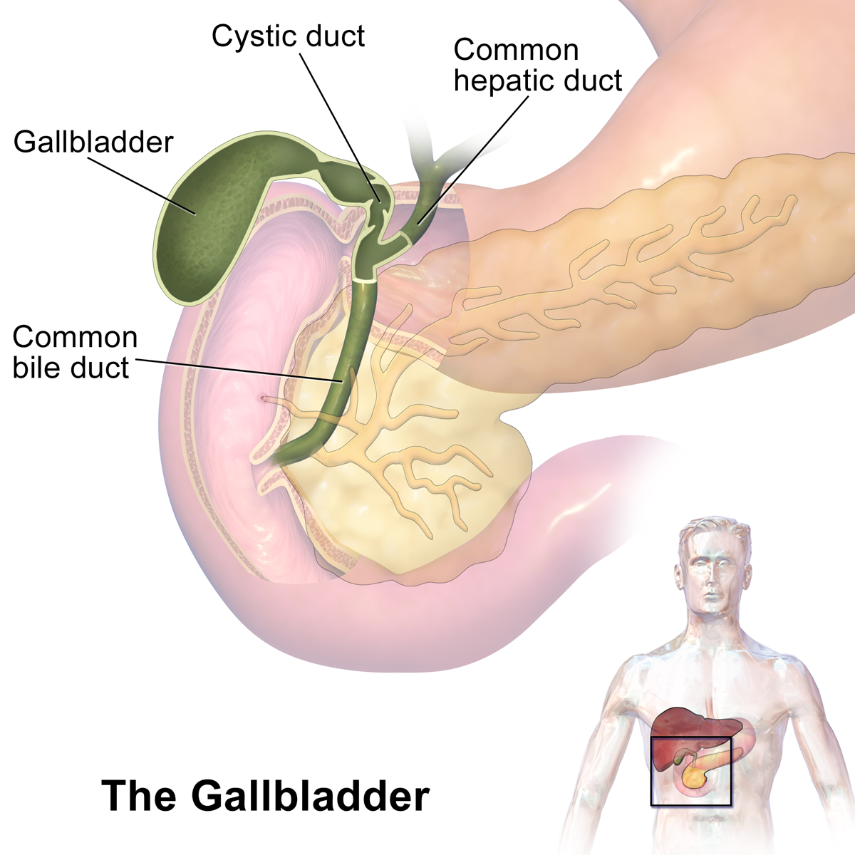


Anatomy and components
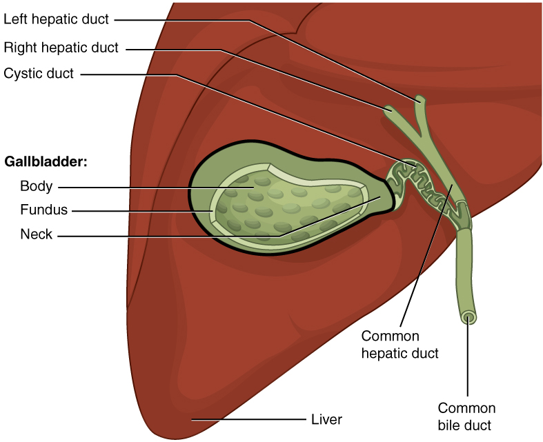
Structurally, the gallbladder has three regions: the fundus (rounded end), the body (central portion), and the neck, which tapers toward the cystic duct. Together the cystic duct and the hepatic ducts form the common bile duct that opens into the duodenum. Typical length varies among individuals, often cited as several centimeters; its internal lining can concentrate bile by absorbing water and electrolytes. For basic information about bile itself, see bile. Average size estimates and measurements are available in standard references (size estimates).
Function in digestion
The gallbladder stores and concentrates bile produced continuously by the liver. When fat enters the small intestine, the hormone cholecystokinin triggers the gallbladder to contract and release bile into the duodenum through the common bile duct. Bile contains bile salts that emulsify dietary fats, aiding enzymes in breaking them down so they can be absorbed. The close anatomical relationship between the gallbladder and the liver and duodenum reflects this coordinated role.
Common conditions and clinical notes
The gallbladder can be affected by several common problems. Gallstones (solid deposits of cholesterol or pigment) are a frequent cause of pain and obstruction. Inflammation of the gallbladder (cholecystitis), infection, and biliary obstruction can produce abdominal pain, fever, or jaundice. Diagnostic methods include ultrasound imaging and blood tests; treatment ranges from diet and observation to surgical removal (cholecystectomy) when complications occur. Typical clinical features and management approaches are described in medical guidelines and patient resources.
Development, variations, and notable facts
Embryologically the gallbladder develops from the foregut and may show anatomical variations in size, shape, or the pattern of ductal connections. Not everyone requires a gallbladder for normal digestion — people can live without one after surgical removal because the liver still secretes bile, although bile is released less rapidly. Historical anatomists and clinicians have studied the biliary system for centuries; modern imaging and surgical techniques have greatly reduced the risks of treatment.
- Major parts: fundus, body, neck, cystic duct, common bile duct.
- Key functions: storage, concentration, regulated release of bile for fat digestion.
- Common problems: gallstones, cholecystitis, biliary obstruction.
For introductory resources and clinical overviews consult reputable medical sites and textbooks (see links for further reading: shape, location, abdominal relations, bile composition, measurements, liver relation, duodenal connection).
Related articles
Author
AlegsaOnline.com Gallbladder — structure, function, and clinical significance Leandro Alegsa
URL: https://en.alegsaonline.com/art/37297