
Smooth muscle: structure, function, locations and clinical relevance
Smooth muscle is non‑striated, involuntary muscle found in organs and vessels. This article explains its structure, types, physiology, locations and clinical significance in health and disease.
Overview
Smooth muscle is the involuntary muscle tissue found in the walls of many internal organs and tubular structures. It differs from skeletal muscle in appearance and control: smooth muscle is not attached to the skeleton and lacks the striated banding seen in voluntary muscle. It generates slow, sustained or rhythmic contractions that control lumen diameter, propel contents along ducts and maintain organ tone. In broad terms, smooth muscle supports the inner workings of the body and contributes to processes ranging from circulation and digestion to vision and reproduction. For a general introduction see internal organs and viscera.
Image gallery
4 Images


Structure and cellular features
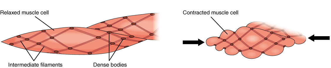
Individual smooth muscle cells are spindle-shaped (fusiform), typically contain a single central nucleus, and range widely in size depending on location and species. The contractile apparatus is composed mainly of actin and myosin but arranged in a less regular lattice than in skeletal muscle; this arrangement explains the non‑striated appearance. Dense bodies and intermediate filaments provide intracellular attachment points for contractile proteins, allowing tension developed within a cell to be transmitted to neighboring cells. Many smooth muscle cells are connected by gap junctions that permit electrical coupling and coordinated contraction across a tissue.
Types and organization
- Single-unit (unitary) smooth muscle: cells are electrically linked and behave as a functional syncytium. This organization produces coordinated waves of contraction such as the peristaltic movements of the gastrointestinal tract or rhythmic activity of the uterus.
- Multi-unit smooth muscle: cells are less coupled electrically and can be activated individually, allowing finer control of tension. Examples include some eye muscles that adjust pupil size and certain portions of the airway.
Mechanisms of contraction and regulation
Contraction is initiated primarily by increases in intracellular calcium concentration, which may arise from entry through membrane channels or release from internal stores. Calcium binds calmodulin and activates myosin light chain kinase, promoting cross-bridge cycling between actin and myosin. Many smooth muscles can enter a low-energy "latch" state in which force is maintained with reduced ATP consumption; this is advantageous for structures that need prolonged tonic contraction, such as some sphincters and blood vessels.
Neural, hormonal and local control
Smooth muscle is regulated by the autonomic nervous system, circulating hormones and local chemical or mechanical signals. Neurotransmitters released by sympathetic and parasympathetic nerves can cause contraction or relaxation depending on the receptor types present. Hormones and paracrine factors (for example nitric oxide, prostaglandins and endothelin) modulate tone and reactivity. Stretch itself can trigger contractile responses in certain tissues, contributing to reflex control of function.
Principal locations and roles
Smooth muscle is widely distributed where controlled constriction, propulsion or regulation of flow is needed. Common locations and their principal roles include:
- walls of blood vessels, where it adjusts lumen diameter and influences blood pressure;
- vessels of the lymphatic system, helping move lymph toward central veins;
- the urinary bladder, enabling storage and coordinated emptying of urine;
- the uterus, whose coordinated contractions are important in childbirth;
- muscular layers of the gastrointestinal tract, producing peristalsis and mixing of contents;
- the respiratory tract, where airway smooth muscle regulates airflow and resistance;
- most exocrine ducts, controlling the passage of secretions;
- specialized structures such as the iris, which adjusts pupil size to light;
- and regions that require fine graded adjustments distinct from skeletal muscle control.
Developmental and molecular markers
During development, smooth muscle cells typically arise from mesenchymal precursors and differentiate under the influence of growth factors and mechanical cues. Molecular markers commonly used to identify smooth muscle include smooth muscle actin and myosin isoforms, though expression patterns vary with tissue type and physiological state. Phenotypic modulation of smooth muscle (shifts between contractile and synthetic states) is a recognized feature in vascular disease and tissue repair.
Clinical relevance and therapeutics
Dysfunction of smooth muscle contributes to a range of disorders. Hyperresponsiveness of airway smooth muscle plays a central role in asthma, while abnormal vascular smooth muscle behavior is implicated in hypertension and atherosclerotic remodeling. Disorders of uterine smooth muscle affect labor, and bladder smooth muscle dysfunction leads to urinary symptoms. Pharmacological agents commonly target smooth muscle: bronchodilators relax airway smooth muscle, vasodilators act on vascular smooth muscle, and antispasmodics reduce unwanted gastrointestinal or urinary contractions. Understanding smooth muscle biology also informs surgical approaches and endovascular therapies.
Research directions and summary
Current research explores smooth muscle signaling pathways, the basis of phenotypic plasticity, and the development of more selective drugs to modulate tone with fewer side effects. Tissue engineering and regenerative medicine efforts aim to restore or replace smooth muscle function in damaged organs. In summary, smooth muscle is a versatile and widely distributed tissue essential to many involuntary functions; its unique cellular organization and regulatory mechanisms allow it to sustain diverse roles in health and disease.
Further reading and resources: general overview, vascular function, lymphatic transport, urodynamic considerations, uterine physiology, airway biology, secretory ducts, ocular mechanics, and comparisons with skeletal muscle.
Questions and answers
Q: What is smooth muscle?
A: Smooth muscle is the type of muscle which is inside the body and not attached to bones.
Q: What controls smooth muscle?
A: Smooth muscle is not under conscious control.
Q: What is the function of smooth muscle?
A: The function of smooth muscle is to help the inner workings of the body.
Q: Where is smooth muscle found?
A: Smooth muscle is found in the walls of blood vessels, vessels of the lymphatic system, the urinary bladder, uterus, male and female reproductive tracts, gastrointestinal tract, respiratory tract, most ducts, and a number of other places such as the iris.
Q: How does the smooth muscle look like?
A: The smooth muscle looks different from the skeletal muscle.
Q: How does smooth muscle work?
A: Smooth muscle can squeeze slowly and move in waves along a duct. Most types of smooth muscle squeeze gently for a long time and use little energy. Some smooth muscle squeezes quickly and relaxes in phases.
Q: What is the difference between smooth muscle and skeletal muscle?
A: Smooth muscle is inside the body, not attached to bones and is not under conscious control, while skeletal muscles are attached to bones and are under conscious control. Additionally, the smooth muscle looks different from the skeletal muscle and works differently.
Related articles
Author
AlegsaOnline.com Smooth muscle: structure, function, locations and clinical relevance Leandro Alegsa
URL: https://en.alegsaonline.com/art/91268