Palatine bone
The palatine bone is a paired L-shaped bone at the back of the nasal cavity and hard palate, contributing to the orbit floor, nasal wall and serving as passage for neurovascular structures.
The palatine bone is a small, paired bone of the facial skeleton that helps form the back part of the hard palate and parts of the nasal cavity. It sits posterior to the maxilla and anterior to the sphenoid bone, and lies on either side of the nose. For a concise definition and diagrams see palatine bone references and resources. It also contributes to the medial floor of the orbit and the lateral wall of the nasal cavity, interfacing with several neighboring bones.
Image gallery
10 Images


Anatomy and parts
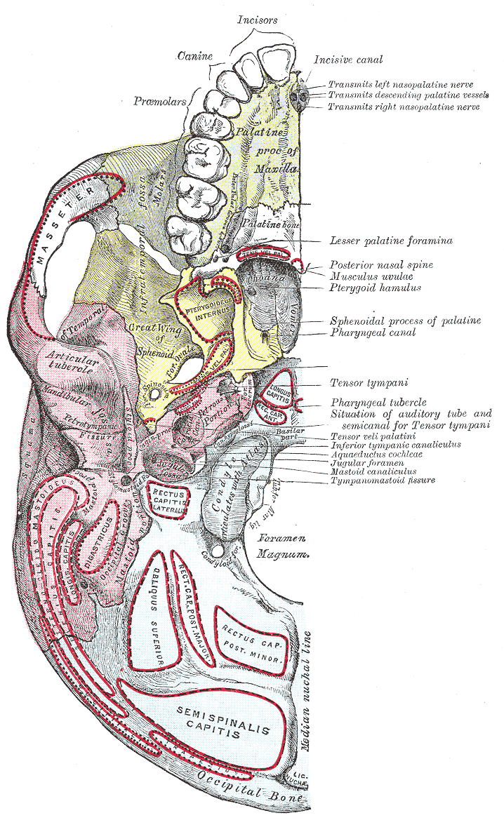
The bone is roughly L-shaped and consists of two main plates: the horizontal (or palatine) plate and the perpendicular (or vertical) plate. Key features include:
- Horizontal plate — forms the posterior portion of the hard palate and meets the opposite palatine bone at the posterior nasal spine.
- Perpendicular plate — contributes to the lateral wall of the nasal cavity and the floor of the orbit.
- Orbital, sphenoidal and pyramidal processes — small projections that contact neighboring bones and complete the facial skeleton.
- Greater and lesser palatine foramina — openings that transmit the palatine nerves and vessels to the palate.
Articulations and relations
The palatine bone articulates with the maxilla, sphenoid, ethmoid, vomer, inferior nasal concha and the opposite palatine bone. Its foramina carry sensory and vascular supply to the hard and soft palate, which is important in dental and ENT procedures.
Development, name and clinical relevance
Ossification of the palatine bones occurs prenatally and they fuse with neighboring bones during growth. The English name derives from Latin palatum (palate). Clinically the palatine bone is significant in cleft palate repair, maxillofacial surgery, and when locating the greater palatine foramen for anesthesia in dentistry. Fractures or congenital malformations can affect speech, swallowing and nasal airflow.
Distinctions frequently noted in anatomical study include the palatine bone versus the nasal cavity structures and the palatine process of the maxilla; the latter is a different bone contribution to the hard palate. For further anatomical images and surgical considerations consult regional anatomy sources and surgical texts linked at clinical references and specialty pages via skeletal overviews or detailed entries.
Related articles
Author
AlegsaOnline.com Palatine bone Leandro Alegsa
URL: https://en.alegsaonline.com/art/74141