
Maxilla (upper jaw bone)
The maxilla is the paired upper jaw bone that forms the central face, supports the upper teeth, helps form the nose and eye sockets, and varies in form and mobility across vertebrates.
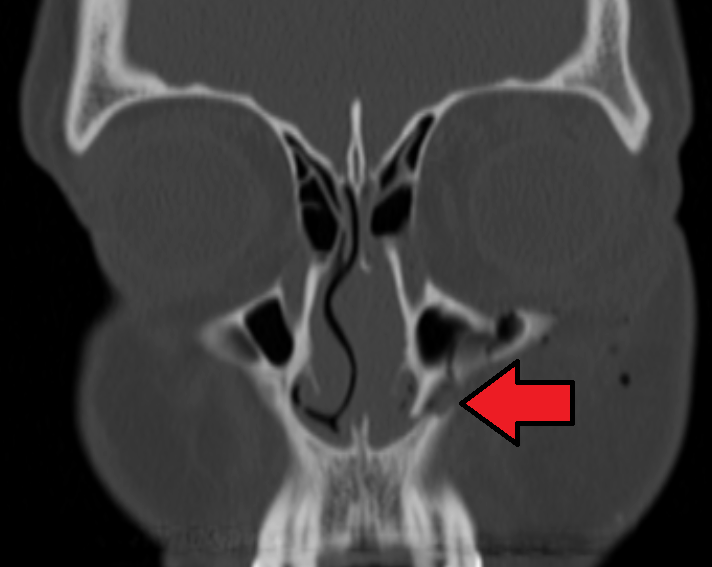
The maxilla, commonly called the upper jaw, is a paired facial bone that forms the middle third of the face. It contributes to the architecture of the oral cavity, the floor of the orbit, the lateral wall and floor of the nasal cavity, and contains the maxillary sinus in mammals. The maxilla bears the upper dentition along its alveolar margin and provides attachment sites for facial muscles and soft tissues.
Image gallery
7 Images




Anatomical parts and characteristics
Each maxilla consists of a central body with several processes that articulate with neighboring bones. Typical features include:
- Body — houses the maxillary (antral) sinus in many mammals and contains the infraorbital canal.
- Alveolar process — the ridge that supports the upper teeth.
- Frontal, zygomatic, palatine and orbital processes — projections that form sutures with the frontal, zygomatic and palatine bones and help make the orbital floor.
- Foramina — openings such as the infraorbital foramen transmit nerves and vessels to the face.
Development and variation
During embryonic development the maxilla arises from the first pharyngeal arch and forms by intramembranous ossification. Growth continues at facial sutures and by remodeling associated with tooth eruption. In humans and most other mammals the paired maxillary bones are firmly joined to surrounding facial bones to form a stable skull. By contrast, in many fish and reptiles the maxilla remains less rigidly attached, allowing greater mobility of the upper jaw during feeding.
Comparative and functional notes
Across vertebrates the maxilla shows important evolutionary differences. In many mammals it is relatively rigid and helps form the oral cavity and sinuses; in various fish and reptiles the bone can be more loosely connected to permit different feeding mechanics. For example, in some snakes the maxilla and other facial bones are highly kinetic, enabling the jaws to move independently and, in extreme cases, to be separated to ingest large prey — a mobility discussed in comparative anatomical literature sources.
Clinical relevance and uses
The maxilla is central to several medical and dental fields. Common clinical considerations include:
- Trauma: fractures of the maxilla (including classic Le Fort patterns) can disrupt occlusion, breathing and eye function.
- Dental care: the alveolar process is important for tooth extraction, implants and orthodontics.
- Surgical procedures: maxillofacial and orthognathic surgeries reshape the maxilla to correct congenital anomalies, deformities, or disease.
- Sinus disease: the proximity of the maxillary sinus makes the bone relevant to ENT and sinus surgery.
Because the maxilla links facial form, oral function and airway anatomy, it is a frequent focus in anatomy education, comparative biology and clinical practice. For accessible overviews, see resources on the upper jaw, vertebrate skull variation such as in fish and reptiles, and specific discussions of mammalian facial anatomy in mammals and snake jaw mechanics in snakes with further reading provided at specialized sources.
Related articles
Author
AlegsaOnline.com Maxilla (upper jaw bone) Leandro Alegsa
URL: https://en.alegsaonline.com/art/63025