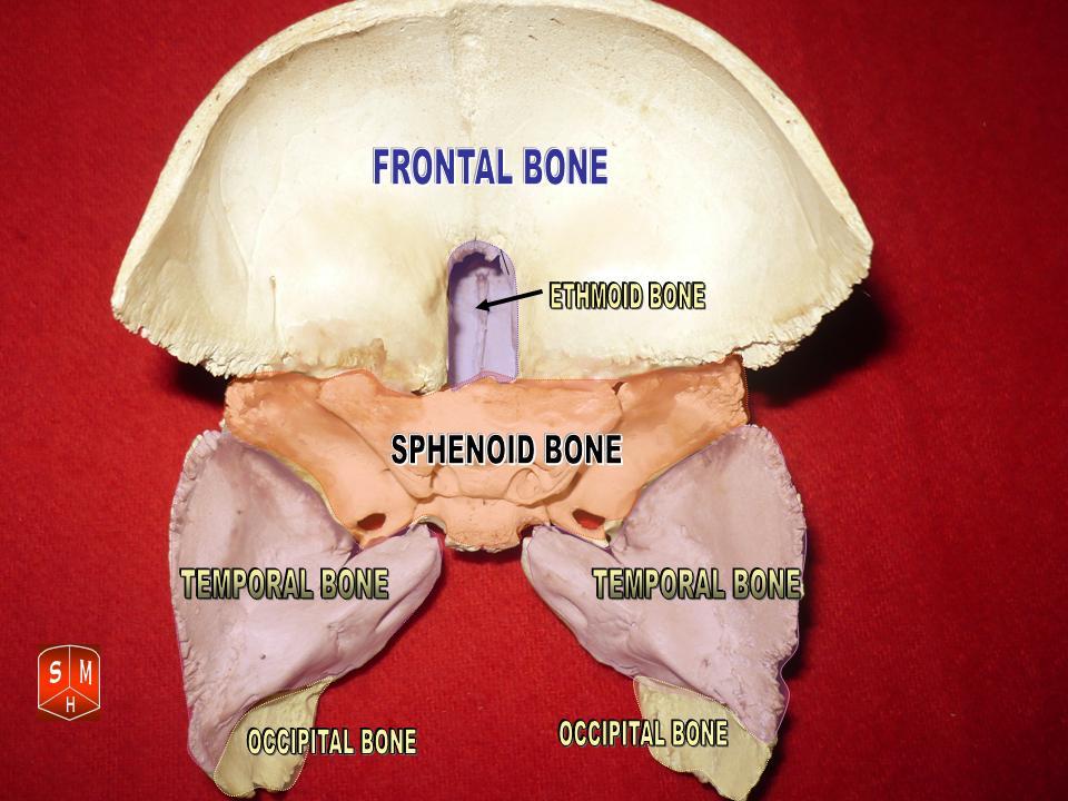
Sphenoid bone
Central unpaired bone of the skull base that helps form the cranial floor, houses the pituitary fossa, contains the sphenoidal sinuses, and transmits several cranial nerves and vessels.
Overview
The sphenoid bone is an unpaired, complex bone situated near the middle of the skull base. It contributes to the floor of the cranial cavity, the lateral walls of the skull, and parts of the orbits. Because of its central position and multiple connections with other cranial and facial bones it is often described as a keystone of the cranial skeleton. For general reference see an anatomical atlas and a concise clinical review.
Image gallery
10 Images


Anatomical parts and key openings
The sphenoid has a central body and several projecting elements. Major components include:
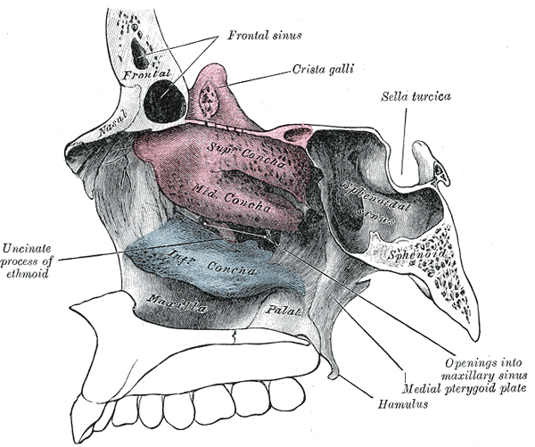
- Body — contains the sphenoidal sinuses and the sella turcica, the depression that houses the pituitary gland.
- Greater wings — extend laterally to form part of the cranial floor and the orbit.
- Lesser wings — form a ridge above the orbit and contribute to the anterior cranial fossa boundary.
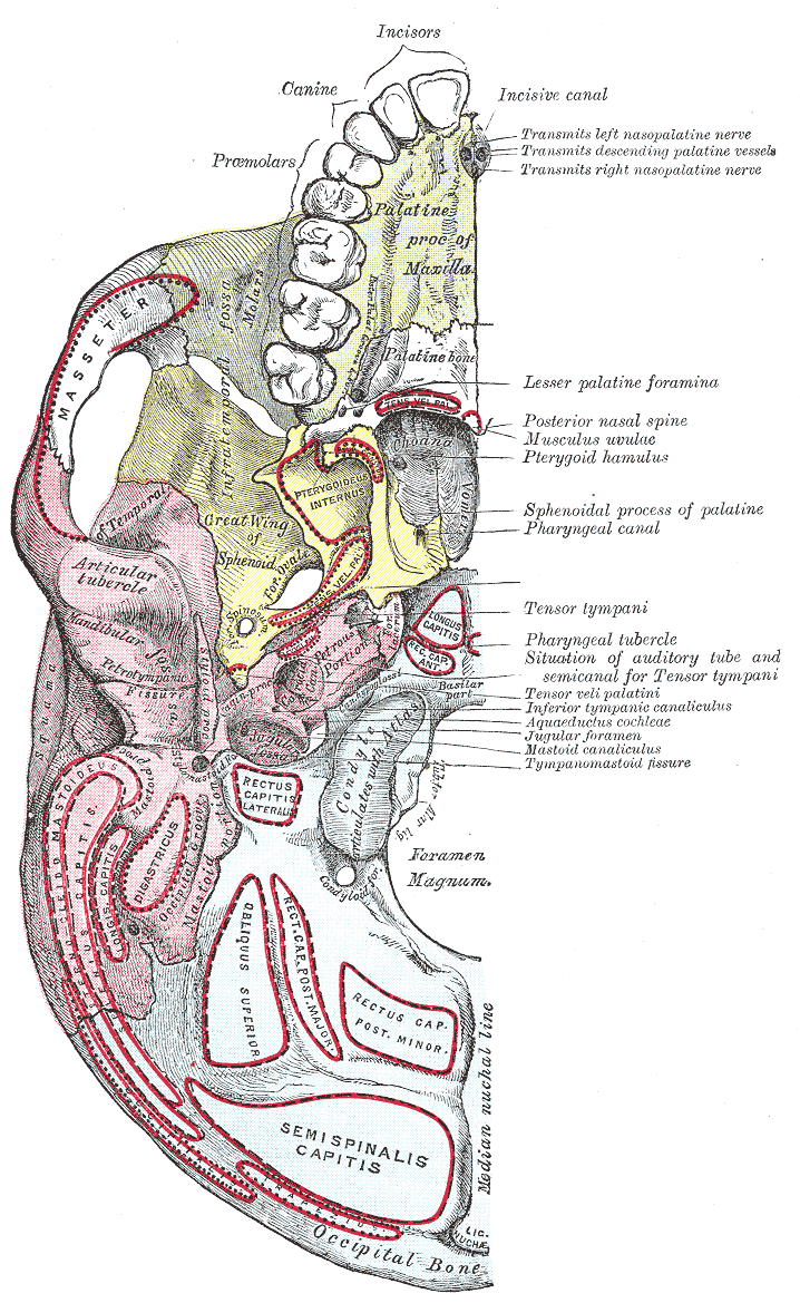
- Pterygoid processes — project inferiorly and give attachment to muscles and fascia of mastication.
Several foramina and fissures in the sphenoid transmit nerves and vessels. Notable openings include the optic canal, superior orbital fissure, foramen rotundum, foramen ovale and foramen spinosum, each serving distinct neurovascular structures.
Development and articulations
The bone develops from multiple ossification centers during fetal and early postnatal life; different parts fuse as the skull matures. The sphenoid articulates with frontal, temporal, parietal, occipital, zygomatic, palatine, vomer and ethmoid bones, linking the facial skeleton to the neurocranium.
Clinical significance
Its central location makes the sphenoid clinically important. The sella turcica is the usual surgical route for pituitary access (transsphenoidal approaches). The sphenoidal sinuses are a site of sinusitis and can be adjacent to critical structures; infections or tumors here may affect cranial nerves or the cavernous sinus. Fractures of the skull base can involve sphenoid foramina and lead to neurovascular injury.
Imaging, variation and notable facts
The sphenoid is readily seen on CT and MRI, which are used to evaluate sinuses, pituitary pathology, fractures and tumor spread. Size and pneumatization of the sphenoidal sinuses vary between individuals; this variability affects surgical planning. As an unpaired, centrally placed bone with complex shape and many foramina, the sphenoid plays a pivotal role in skull anatomy and neurosurgical approaches.
Related articles
Author
AlegsaOnline.com Sphenoid bone Leandro Alegsa
URL: https://en.alegsaonline.com/art/92639
Sources
- merriam-webster.com : Entry "sphenoid"
- merriam-webster.com : Merriam-Webster Online Dictionary