Micrograph: Photomicrography and Microscopic Imaging
A micrograph is a photograph made through a microscope that reveals magnified structure. This article covers types (light, SEM, TEM), preparation, applications, history and good-practice considerations.
Overview
A micrograph (also called a photomicrograph) is a photograph taken through a microscope to record the appearance of small structures at magnified scale. Micrographs translate features that are invisible to the naked eye into images that can be examined, measured, compared, and published. They are essential tools across biology, materials science, geology, electronics, and forensic analysis.
Image gallery
9 Images


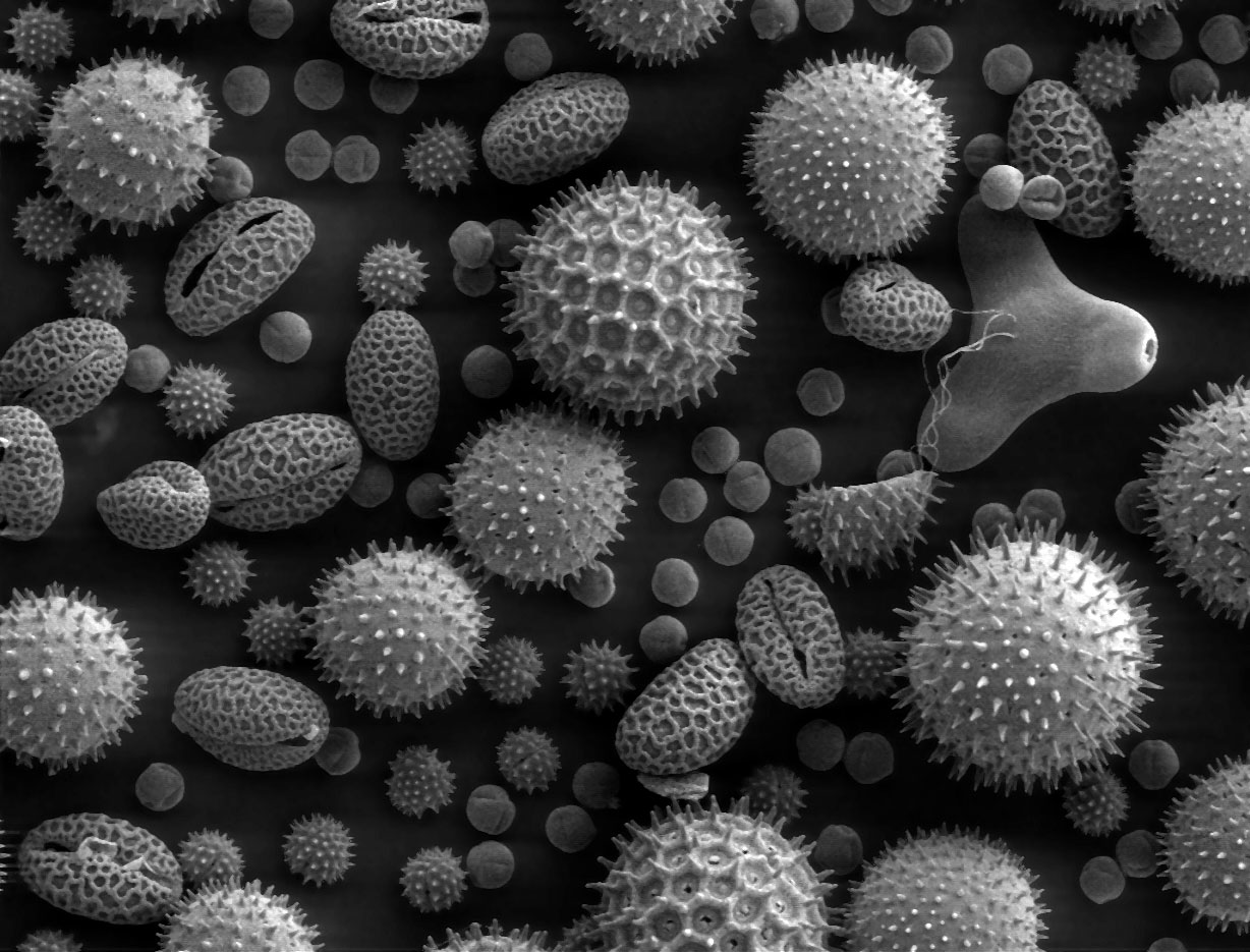


Types and characteristics
Micrographs differ by the imaging modality and the information they provide. Common types include optical (light) micrographs, produced with compound microscopes using transmitted or reflected light; scanning electron micrographs (SEM), which show surface topography and composition contrast; and transmission electron micrographs (TEM), which reveal internal ultrastructure at much higher resolution. Optical micrographs may be in color; electron micrographs are typically grayscale and are often presented with false color to emphasize features. For portable background reading on microscopes, see light microscopy resources.
Preparation and imaging techniques
Preparation depends on the specimen and the imaging method. Biological samples often require fixation and staining to enhance contrast; thin sectioning is needed for TEM; nonconductive materials for SEM are usually coated with a thin conductive layer. Imaging techniques include brightfield, darkfield, phase contrast and differential interference contrast for light microscopy, and secondary/backscattered electron imaging for SEM. Proper calibration, inclusion of a scale bar, and metadata are critical so magnification and measurements remain meaningful.
Uses and examples
Micrographs support a wide range of applications: documenting cells and tissues in medical research; identifying minerals and textures in petrography; analyzing fracture surfaces in forensic engineering (forensic engineering) and trace evidence in forensic science (forensic science); inspecting microelectronic structures; and selecting areas for more detailed analysis during scanning electron microscopy. In education, micrographs help teach structure–function relationships by making the invisible visible.
History, standards and interpretation
Photomicrography evolved in the 19th century with the development of practical microscopes and photographic techniques. Since then, standards for image acquisition and reporting have grown: manuscripts and technical reports generally require scale bars, resolution statements, and disclosure of any digital enhancement to avoid misleading interpretation. Users should distinguish between magnification (a display property) and resolution (an intrinsic limit set by the method and instrument), noting that light microscopes are limited by the wavelength of light while electron microscopes can resolve much smaller features.
Best practices and notable distinctions
- Always include a calibrated scale bar and instrument settings when possible.
- Avoid excessive post-processing that alters scientific meaning.
- Use the appropriate modality: choose SEM for surface detail, TEM for internal ultrastructure, and light microscopy for live-cell imaging and colored stains.
Well-prepared micrographs are powerful records that combine careful physical preparation, appropriate imaging choices, and transparent reporting to communicate microscopic observations reliably to other researchers and practitioners.
Related articles
Author
AlegsaOnline.com Micrograph: Photomicrography and Microscopic Imaging Leandro Alegsa
URL: https://en.alegsaonline.com/art/64600