
Femoral artery: anatomy, function, and clinical importance
Major artery supplying the lower limb; origin, course, branches, surface landmarks, clinical uses and common problems including access for procedures and trauma management.
The femoral artery is the principal arterial supply to the lower limb. It is the anatomical continuation of the external iliac artery as it passes under the inguinal ligament. The vessel delivers oxygen-rich blood to tissues of the thigh, leg and foot and is both a useful surface landmark and a frequent site for medical access.
Image gallery
10 Images


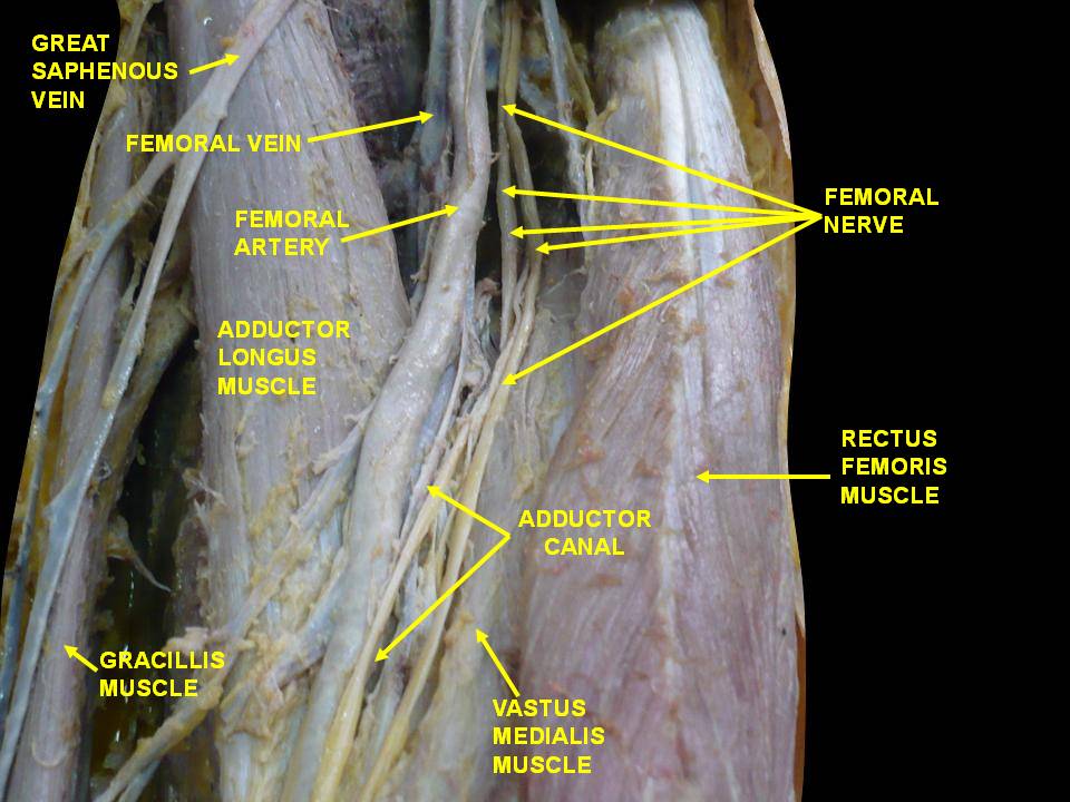

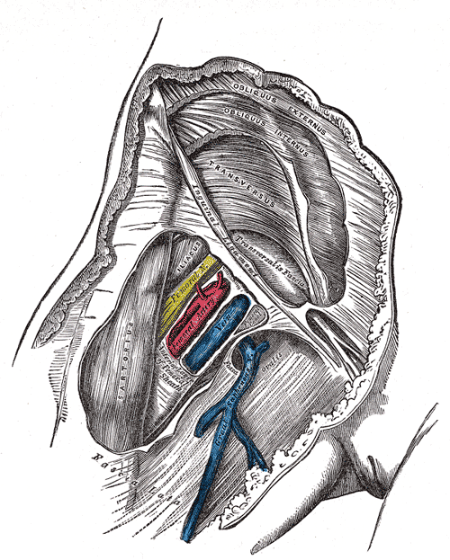
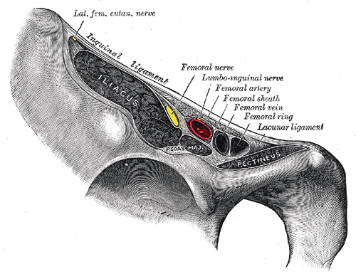
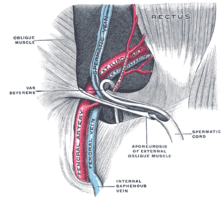
Anatomy and course
After the external iliac artery crosses beneath the inguinal ligament it becomes the femoral artery. It travels down the front of the thigh within the femoral triangle, then continues more deeply and ultimately becomes the popliteal artery behind the knee when it passes through the adductor (Hunter's) canal. Major branches include the profunda femoris (deep femoral) artery and several perforating branches that supply the thigh muscles; the superficial femoral artery is the term commonly used for the main downward continuation.
Relations and surface landmarks
In the groin the artery lies relatively close to the skin, immediately lateral to the femoral vein and medial to the femoral nerve in the region often remembered by the mnemonic NAVEL (Nerve, Artery, Vein, Empty space, Lymphatics from lateral to medial). Its position under the inguinal ligament makes the groin a convenient site to palpate the femoral pulse and to apply direct pressure for hemorrhage control.
Function and clinical uses
The femoral artery supplies arterial blood to the lower extremity and contributes to collateral circulation around the hip and thigh. Clinically it is an important access site for vascular procedures: catheterization, angiography, percutaneous coronary interventions and endovascular repairs may be performed via femoral arterial puncture. Surgeons also use it for bypass graft inflow or for direct arterial cannulation during resuscitation.
- Common clinical examinations: checking the femoral pulse, assessing peripheral circulation.
- Procedures: arterial access for angiography, endovascular interventions, and intra-arterial monitoring.
- Emergency care: compression or surgical control to manage life-threatening groin hemorrhage.
Disorders and significance
The femoral artery can be affected by atherosclerotic narrowing, aneurysm formation, traumatic injury and iatrogenic complications from catheterization (such as hematoma or pseudoaneurysm). Because of its superficial location in the groin, the artery is vulnerable to penetrating trauma but also accessible for lifesaving interventions and diagnostic sampling.
For general information consult anatomical texts or clinical guidelines: see resources on arterial anatomy and vascular access. Additional material is available on the external iliac origin and groin anatomy via cardiovascular sources, on oxygen delivery and blood flow via general physiology, and procedural guidance via interventional references. Practical anatomical descriptions and palpation landmarks are discussed in regional anatomy guides (groin anatomy) and vascular surgery references (femoral artery overview).
Historically the name femoral derives from its relationship to the femur (thigh bone). Understanding its course, branches and surface relationships is essential for clinicians in diagnostics, surgery and emergency care.
Related articles
Author
AlegsaOnline.com Femoral artery: anatomy, function, and clinical importance Leandro Alegsa
URL: https://en.alegsaonline.com/art/33961
Sources
- wheelessonline.com : "Femoral Artery"