
Artery: structure, function, and clinical importance
Arteries are blood vessels that carry blood away from the heart. This article explains their structure, types, function, fetal exceptions, and common clinical conditions in clear, neutral terms.
Overview
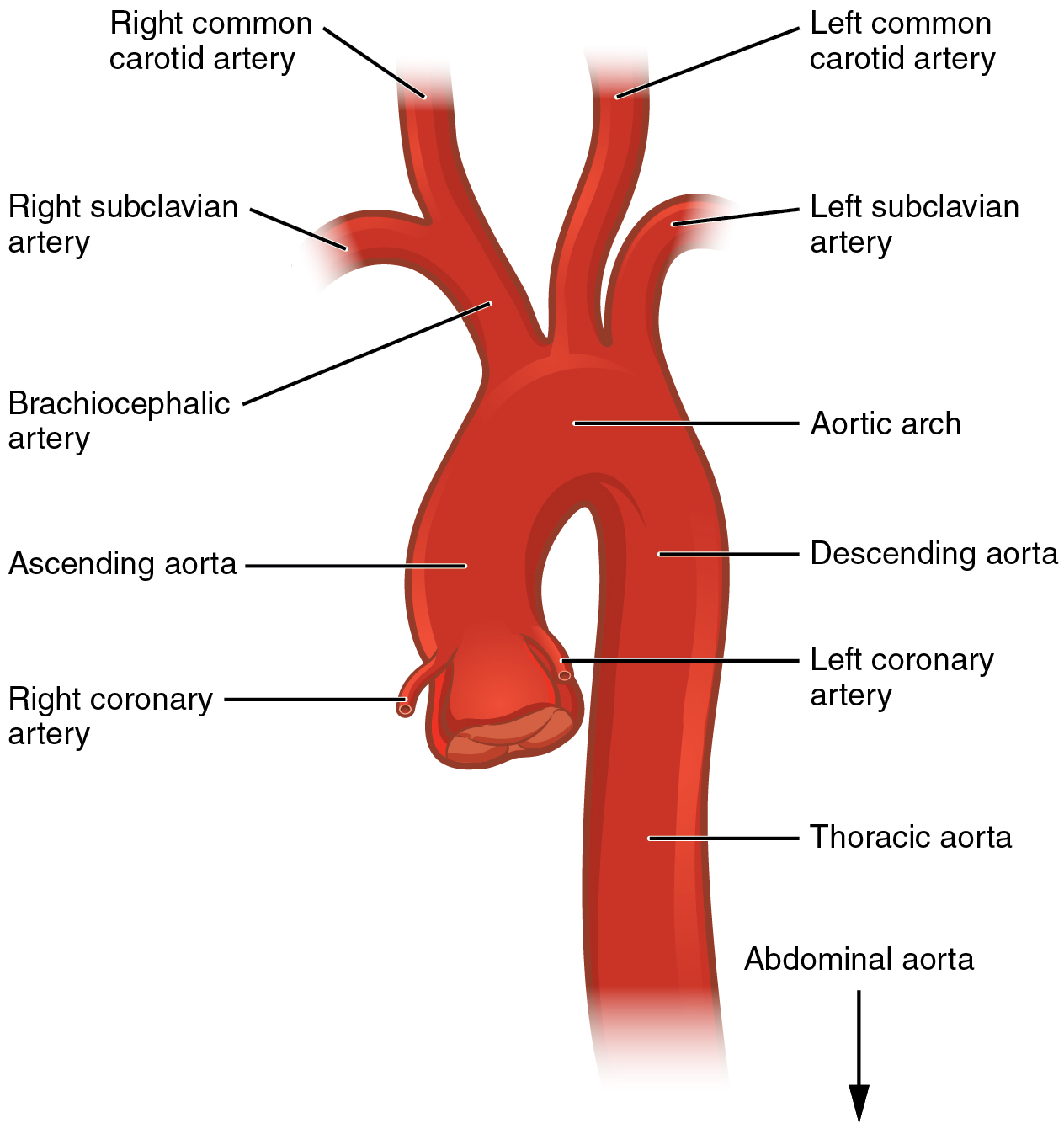
An artery is a type of blood vessel whose principal role is to carry blood away from the heart toward organs and tissues. In most animals, arteries transport oxygenated blood; notable exceptions are the pulmonary artery, which carries blood to the lungs for oxygenation, and the umbilical artery in the fetus, which returns deoxygenated blood to the placenta.
Image gallery
4 Images


Structure and layers
Arteries have a layered wall that supports their high-pressure role. From outside in these are the adventitia (outer layer), the media (muscular middle layer) and the intima (inner lining). The outer coat contains connective and stretchy tissue that resists tearing. The middle layer is rich in smooth muscle and elastic fibers so the vessel can widen (dilate) or narrow (constrict) to regulate flow. The inner surface is a single-cell lining of endothelium, the same cells that line the heart chambers, which provides a smooth barrier and participates in local signaling.
Types and branching
Large arteries near the heart are often elastic to accommodate the pulse of blood; farther from the heart arteries become more muscular and control distribution. Arteries progressively branch into smaller vessels called arterioles, which in turn feed capillary networks where exchange with tissue occurs.
Function and distinguishing features
Because arterial pressure follows heart contractions, the pulse and much of the circulating pressure are borne by arteries. They are generally located deep under the skin, unlike many veins. Most arteries do not contain valves; instead, their elastic recoil and muscular tone help propel blood onward between heartbeats. Clinically, arteries are used for measuring blood pressure and for sampling arterial blood gases.
Clinical relevance and examples
- Atherosclerosis: buildup of plaques in arterial walls that can reduce flow and lead to heart attack or stroke.
- Aneurysm: a focal dilation of an artery that can risk rupture if it enlarges.
- Occlusion and ischemia: sudden blockage of an artery can cause tissue damage due to lack of oxygen.
Historical and developmental notes
The basic arterial plan is conserved across vertebrates, adapted in size and elasticity to an animal's physiology. During development some vessels have temporary roles (for example the fetal umbilical artery and other shunts); after birth many of these pathways close as the lungs and systemic circulation become dominant.
Key characteristics
- Carry blood away from the heart (heart, vessel).
- Three-layered wall with elastic and muscular components (stretchy tissue, widen/narrow).
- Generally oxygen-rich except pulmonary and fetal examples (pulmonary artery, fetus).
- Do not typically have valves; pulse and recoil aid forward flow (endothelium, location).
For further reading and visual diagrams see resources indicated by the links above or consult clinical texts about cardiovascular anatomy and disease.
Questions and answers
Q: What is an artery?
A: An artery is a blood vessel that carries blood away from the heart to other parts of the body.
Q: What type of blood do almost all arteries carry?
A: Almost all arteries carry blood with a lot of oxygen in it.
Q: Which arteries do not carry oxygen-rich blood?
A: The pulmonary artery, and (in a fetus) the umbilical artery do not carry oxygen-rich blood.
Q: What are the three layers of an artery?
A: Every artery has three layers: the outer layer is a thick covering made of stretchy tissue, the middle layer is made of muscle, and the inner lining is made of the same cells that line the heart.
Q: Do arteries have valves to prevent blood from flowing backwards?
A: Arteries do not have valves to prevent blood from flowing backwards.
Q: How do arteries push blood through?
A: Every time the heart beats, the arteries stretch out so the blood has room to go through. Once the walls have completely stretched out, the muscles in the arteries make them shrink back to their normal size, pushing the blood through.
Q: What are arterioles?
A: Arteries split down into small vessels called arterioles.
Tags
Related articles
Author
AlegsaOnline.com Artery: structure, function, and clinical importance Leandro Alegsa
URL: https://en.alegsaonline.com/art/6252
Sources
- britannica.com : "Artery: Anatomy"
- cedars-sinai.edu : "Anatomy of the Aorta and Heart"
- brooksidepress.org : "U.S. Navy Standard First Aid Manual, Chapter 3: Bleeding"
- whqlibdoc.who.int : Global atlas on cardiovascular disease prevention and control
- britannica.com : "Aneurysm: Pathology"