Seminiferous tubule — structure, function and clinical significance
Seminiferous tubules are coiled tubes in the testes where sperm form. This article explains their anatomy, cell types, role in spermatogenesis, hormonal control, and clinical conditions that affect fertility.
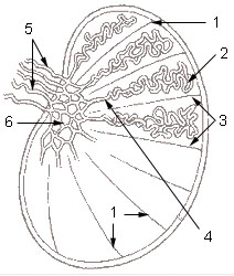
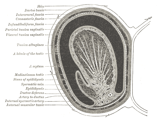
Overview: Seminiferous tubules are highly coiled channels within each testis that serve as the primary site of sperm production. Their intricate internal lining supports development of male germ cells into mature sperm while separating the process from systemic circulation.
Image gallery
6 Images


Structure and main cell types
The tubules are bounded by a basement membrane and layers of contractile peritubular cells. The germinal epithelium lining the lumen contains developing germ cells at successive stages and specialized supporting cells called Sertoli cells. Interstitial tissue between tubules contains Leydig cells that produce testosterone.
Function and physiology
Spermatogenesis is a coordinated series of cell divisions and maturation within the tubules. Sertoli cells provide physical support, nutrients, phagocytose residual bodies and form tight junctions that create the blood–testis barrier. Hormones such as follicle-stimulating hormone and intratesticular testosterone regulate the process.
Key stages and components
- Spermatogonial proliferation
- Meiotic division to reduce chromosome number
- Spermiogenesis: morphological transformation into motile sperm
Clinical significance: Damage to seminiferous tubules—by infection, heat, toxins, obstruction or vascular problems—can impair sperm production and fertility. Diagnostic evaluation may include biopsy and imaging. Understanding tubule histology is important in reproductive medicine.
Distinctions: Seminiferous tubules differ from the ductal system (rete testis and epididymis) that stores and transports sperm after they leave the tubules. Their role is primarily generative rather than storage or maturation outside the testis.
Related articles
Author
AlegsaOnline.com Seminiferous tubule — structure, function and clinical significance Leandro Alegsa
URL: https://en.alegsaonline.com/art/88751