Serosa (serous membrane): structure, function, and clinical relevance
A serosa is a thin serous membrane of mesothelial cells and connective tissue that lines closed body cavities (pleura, pericardium, peritoneum), reducing friction and participating in fluid exchange.
Overview
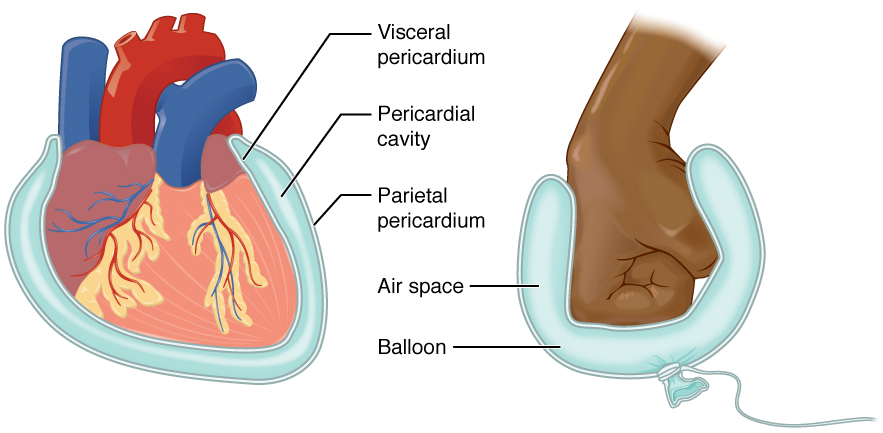
The serosa, also called a serous membrane, is a smooth tissue layer that lines the inner surfaces of closed body cavities and covers many internal organs. Composed of a simple squamous epithelial layer overlying a thin connective tissue bed, the serosa secretes a small amount of lubricating fluid to allow adjacent structures to glide with minimal friction. Major examples are the pleura (around the lungs), the pericardium (around the heart) and the peritoneum (lining the abdominal cavity).
Image gallery
4 Images

Structure and components
Microscopically the epithelial component of the serosa is called the mesothelium, a single layer of flattened cells specialized for fluid transport and surface lubrication. Beneath the mesothelium lies a delicate layer of connective tissue that contains small blood vessels, lymphatics and nerves. Together these layers form a thin, flexible covering that tolerates stretching and sliding. The serous fluid it produces is a clear, protein-poor exudate that reduces friction during movement of organs.
Locations and examples
Serosae are found wherever organs move within closed cavities. Common named serous membranes include:
- Pleura — the paired membranes surrounding the lungs and lining the thoracic cavity.
- Pericardium — the double-layered sac around the heart.
- Peritoneum — the membrane lining the abdominal cavity and covering many visceral surfaces.
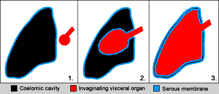
Some structures that lie outside these closed cavities are instead bound by connective tissue called adventitia. For example, the majority of the esophagus and the distal rectum lack a serosal covering and are described as having an adventitial surface. The serosa typically seals or reflects around movable organs within the abdominal cavity, creating potential spaces and fluid compartments.
Functions and clinical importance
Primary functions of the serosa include mechanical protection, facilitation of organ movement, limited fluid exchange, and a role in local immune responses. Clinically, serosal surfaces are central to several common conditions and procedures. Inflammation of a serous membrane—pleurisy, pericarditis or peritonitis—produces pain and can generate excess fluid (effusion or ascites). Malignancies such as mesothelioma arise from mesothelial cells. The peritoneum is also exploited therapeutically in peritoneal dialysis because of its large surface area for solute and fluid exchange.
Development, distinctions and notable facts
Embryologically, serosal linings derive from the mesoderm (specifically lateral plate mesoderm), which differentiates into parietal and visceral mesothelial layers during body-cavity formation. Important distinctions for anatomy and surgery are the difference between serosa and adventitia (sliding lubrication versus firm attachment), and between serosa and mucosa (serosa covers closed cavities; mucosa lines open alimentary and respiratory tracts). Surgeons exploit serosal planes to mobilize organs, and pathologists examine serosal surfaces for signs of spread of infection or malignancy.
Common conditions and examples
- Pleuritis (pleurisy) — painful inflammation of the pleura, sometimes with pleural effusion.
- Pericarditis — inflammation of the pericardial serosa, which can impair cardiac function.
- Peritonitis and ascites — infection or fluid accumulation in the peritoneal cavity.
- Mesothelioma — a rare cancer of mesothelial cells, most often linked to asbestos exposure.
Understanding serosal anatomy helps explain patterns of disease spread, approaches to abdominal and thoracic surgery, and the principles behind certain therapeutic procedures. For further fundamentals on cell types and cavity organization, consult standard anatomy references or pathology overviews.
Questions and answers
Q: What is serosa?
A: Serosa is a smooth membrane that consists of a thin layer of cells and a thin connective tissue layer, found on the outer wall of the organs of the abdominal cavity known as the serous cavity.
Q: What does serosa secrete?
A: Serosa secretes serous fluid which reduces friction from muscle movement.
Q: What is the difference between serosa and adventitia?
A: Serosa reduces friction between structures while adventitia binds the structures together.
Q: What is pericardium?
A: Pericardium is the serous membrane covering the heart and lining the mediastinum.
Q: What is pleura?
A: Pleura is the serous membrane lining the thoracic cavity and surrounding the lungs.
Q: What is peritoneum?
A: Peritoneum is the serous membrane lining the abdominopelvic cavity and the viscera.
Q: Which parts of the gastrointestinal tract do not have serosa?
A: The esophagus, as well as the middle and distal rectum, do not have serosa.
Related articles
Author
AlegsaOnline.com Serosa (serous membrane): structure, function, and clinical relevance Leandro Alegsa
URL: https://en.alegsaonline.com/art/89082