Sertoli cell: supporting cells of the testis and guardians of spermatogenesis
Sertoli cells are somatic cells in seminiferous tubules that nurture developing germ cells, form the blood–testis barrier, secrete regulatory factors and maintain the stem cell niche for sperm production.
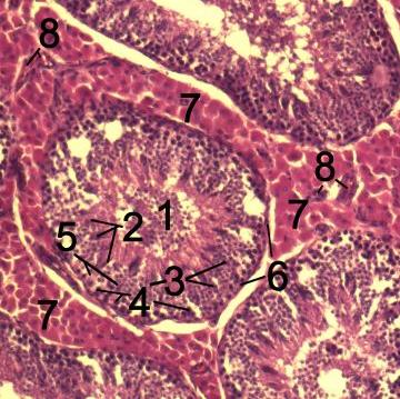
Sertoli cells are specialized somatic cells located within the seminiferous tubules of the testis. Often called "nurse" or "mother" cells, they provide physical support, nutrients and regulatory signals that guide male germ cells through the stages of spermatogenesis, from spermatogonial stem cells through meiosis to differentiate into mature sperm. Their extensive cytoplasmic processes contact multiple germ cells at different stages, creating a coordinated microenvironment essential for orderly development.
Image gallery
2 Images
Characteristics and principal functions
Sertoli cells extend from the basement membrane to the lumen of the seminiferous tubule. They form specialized intercellular junctions, including tight junctions that establish the blood–testis barrier. This barrier divides the seminiferous epithelium into basal and adluminal compartments, regulates passage of molecules and protects developing germ cells from harmful substances and immune attack.
- Structural support and guidance: Sertoli cells physically organize germ cells and help coordinate mitotic and meiotic divisions as well as spermiogenesis.
- Secretion of regulatory proteins: they produce androgen-binding protein (ABP), inhibin B and growth factors that influence local germ cell development and feedback on the pituitary–testis axis.
- Phagocytosis and clearance: they remove residual cytoplasm from late spermatids and clear apoptotic germ cells, maintaining tubule health.
- Immune privilege and barrier function: by contributing to immune modulation within the testis, Sertoli cells help prevent autoimmune reactions against germ cell antigens.
Development and hormonal regulation
Sertoli cells differentiate early in fetal testis development under genetic signals that include the action of SRY. In the embryo they secrete anti-Müllerian hormone (AMH), which promotes regression of the Müllerian ducts and is important for male patterning. In postnatal life and adulthood, Sertoli cell function is regulated primarily by follicle-stimulating hormone (FSH) and by testosterone acting via the androgen receptor; these hormones modulate Sertoli cell secretory activity, the supportive microenvironment and the stem cell niche for ongoing sperm production.
Clinical significance and research
Dysfunction of Sertoli cells can impair spermatogenesis and contribute to male infertility. A well-known condition, Sertoli cell‑only syndrome, describes seminiferous tubules that lack germ cells and is a cause of non‑obstructive azoospermia. Levels of Sertoli cell markers such as inhibin B and AMH are used clinically to assess testicular function in certain settings. Rarely, Sertoli cell tumors arise as sex cord‑stromal neoplasms and may present with endocrine effects.
Because of their role in immune regulation and support of germ cells, Sertoli cells are the subject of research into male contraception, fertility preservation, in vitro models of spermatogenesis and experimental approaches to immunoprotective cell transplantation. Studies of Sertoli cell biology continue to inform understanding of the stem cell niche, hormonal regulation of gametogenesis and potential therapies for male reproductive disorders.
Historical note: The cell type is named after Enrico Sertoli, the 19th‑century Italian anatomist who first described these supportive cells. Today they are recognized as central organizers of the seminiferous epithelium and indispensable for continuous sperm production.
Related articles
Author
AlegsaOnline.com Sertoli cell: supporting cells of the testis and guardians of spermatogenesis Leandro Alegsa
URL: https://en.alegsaonline.com/art/89105