Pons: structure, function, development and clinical significance
The pons is the bulbous part of the brainstem that links the cerebrum, cerebellum and medulla. It contains nuclei, fibre tracts and reticular networks essential for motor control, sensation, breathing and sleep.
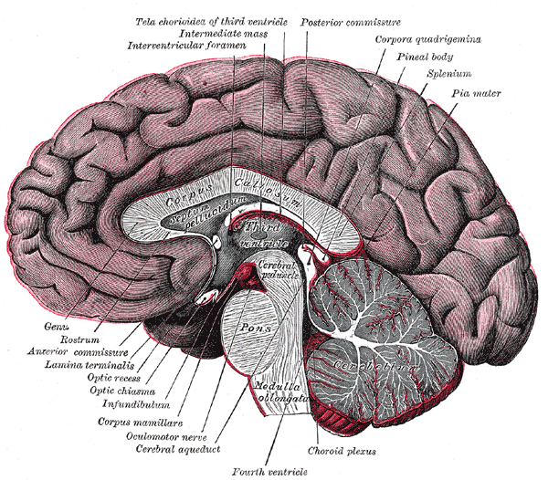
The pons is a prominent bulge on the brainstem that lies between the midbrain above and the medulla oblongata below. It is part of the posterior brain known as the hindbrain and forms a major bridge of nerve fibres connecting higher centres to the cerebellum and spinal cord. Superficially it appears as a band of white matter interspersed with islands of nuclei that perform specialised functions.
Image gallery
7 Images


Anatomy and internal organization
Anatomically the pons is divided into a ventral basal portion (basis pontis) and a dorsal tegmental region. The ventral area contains descending motor tracts and pontine nuclei; the dorsal tegmentum contains cranial nerve nuclei, the reticular formation, and ascending sensory tracts. Massive transverse fibres, the pontocerebellar fibres, sweep laterally to form the middle cerebellar peduncles and link the pons with the cerebellum. Within its core lie both white matter pathways and collections of gray matter that relay and process information.
Functions
The pons acts chiefly as a conduit and a processor. Descending fibres carry motor commands from the cerebral cortex and cerebrum toward the medulla and spinal cord, while ascending tracts transmit sensory information upward to the thalamus. Embedded nuclei contribute to facial sensation and movement, hearing and balance, eye movements and autonomic functions. It also hosts components of respiratory control, elements of sleep regulation (notably REM sleep mechanisms), and part of the reticular activating system that influences arousal.
Cranial nerve relations and notable features
- The pons contains nuclei associated with cranial nerves V (trigeminal), VI (abducens), VII (facial) and VIII (vestibulocochlear).
- Its pontine nuclei form a major relay to the cerebellum via the middle cerebellar peduncle, essential for coordinated movement.
- It comprises both gray nuclei and long white matter tracts often referred to as white fibres in anatomical descriptions.
Because of its central location, small lesions in the pons can produce profound deficits in motor control, cranial nerve function or consciousness.
Development, evolution and clinical importance
Embryologically the pons arises from the metencephalon, the rostral subdivision of the hindbrain, growing alongside the developing cerebellum. It is conserved across vertebrates as a major hub for sensorimotor integration. Clinically, pontine damage can result from stroke, haemorrhage, demyelination (for example central pontine myelinolysis), infection or tumours. Severe ventral pontine injuries may interrupt corticospinal and corticobulbar tracts and cause syndromes characterized by paralysis with preserved consciousness. Neuroimaging commonly examines the pons when patients present with altered breathing, facial weakness, double vision or sudden changes in balance.
In neuroscience research the pons remains important both as a structural relay and as a locus for studying sleep circuits, respiratory rhythm generation and cerebellar communication. For concise anatomical summaries and images consult standard neuroanatomy texts and online resources via the linked entries: brain, hindbrain, medulla oblongata, midbrain, and specialized pages on fibre systems and thalamic relays: white fibres and thalamus.
Understanding the pons helps explain how the nervous system integrates voluntary movement, reflexes and vital functions through an organised mix of nuclei and long-range pathways, serving as a true bridge between the forebrain, cerebellum and spinal cord.
Related articles
Author
AlegsaOnline.com Pons: structure, function, development and clinical significance Leandro Alegsa
URL: https://en.alegsaonline.com/art/77939