Cerebellum: structure, function, development, and clinical significance
The cerebellum is a major brain region that coordinates movement, balance, posture and contributes to motor learning and cognitive tasks. This entry explains its anatomy, connections, functions and clinical relevance.
The cerebellum is a paired, densely folded part of the hindbrain that plays a central role in coordinating voluntary movement and maintaining equilibrium. Found across vertebrates, it sits beneath the cerebral hemispheres and behind the brainstem and is sometimes described as a separate lobe of the brain. Although smaller than the cerebrum by volume, the cerebellum contains a very large proportion of the brain's neurons and a characteristic layered cortex.
Image gallery
10 Images


Structure and components
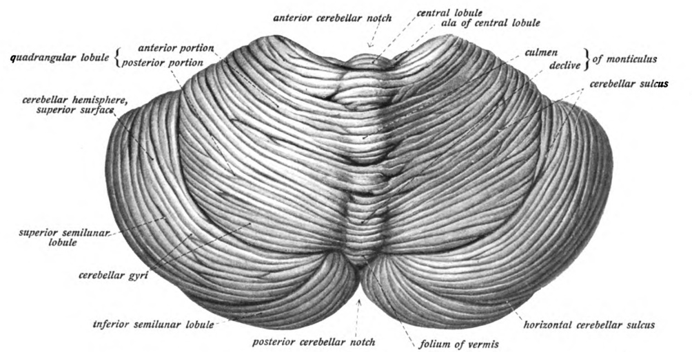
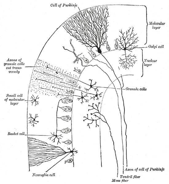
Grossly the cerebellum consists of a central vermis and two lateral hemispheres, and is divided into lobes (anterior, posterior and flocculonodular). Its surface shows folia—narrow, leaf‑like folds. Internally it contains a thin cortical sheet of three layers, large output neurons called Purkinje cells, and deep cerebellar nuclei such as the dentate and fastigial nuclei. Long bundles of axons form pathways that link the cerebellum with the brainstem and spinal cord and are grouped into three cerebellar peduncles.
Connections and pathways
Inputs come from the spinal cord, vestibular system, and cerebral cortex through mossy and climbing fibers; outputs originate from Purkinje cells that inhibit the deep nuclei, which then project to motor and premotor centers via the thalamus and brainstem. The cerebellum communicates extensively with the pons—pontine fibers entering through the middle peduncle convey cortical information to the cerebellum, and reciprocal links with the pons and other brainstem nuclei are crucial for sensorimotor integration.
Functions and importance
Classically the cerebellum refines movement: it times muscle activation, adjusts force and trajectory, and helps keep balance and posture. It is essential for motor learning—acquiring and fine‑tuning skills like riding a bicycle or playing an instrument—by comparing intended actions with sensory feedback and updating future commands. In humans, growing evidence also implicates cerebellar regions in language, attention, working memory and some aspects of social cognition, though these nonmotor roles use circuits distinct from those that control limbs.
Development and evolution
Developmentally the cerebellum arises from the embryonic rhombic lip and metencephalon, and its layered cortex forms through organized migration of neurons. Evolutionarily it is present across jawed vertebrates; its size and complexity vary by species, being particularly elaborate in birds and mammals where precise sensorimotor control and learned behaviors are prominent.
Clinical relevance
Damage or degeneration of the cerebellum produces characteristic signs: ataxia (incoordination), dysmetria (misjudged distances), intention tremor, nystagmus and hypotonia. Causes include stroke, tumors, genetic ataxias, chronic alcohol exposure and autoimmune or paraneoplastic processes. Clinical examination, imaging and genetic testing help localize lesions and guide management. Rehabilitation and targeted therapies can partially restore function because the cerebellum is involved in motor relearning.
- Major components: vermis, hemispheres, lobes, folia, Purkinje cells, deep nuclei
- Key functions: coordination, balance, motor learning, timing, cognitive support
- Common problems: ataxia, dysarthria, nystagmus, tremor
For further anatomical images and functional studies consult specialist neuroanatomy and neuroscience resources or review articles on cerebellar circuits and clinical syndromes (vertebrate examples, brain overviews, fiber anatomy via axonal pathways and pontine relations at pons).
Related articles
Author
AlegsaOnline.com Cerebellum: structure, function, development, and clinical significance Leandro Alegsa
URL: https://en.alegsaonline.com/art/18168