
Occipital bone — anatomy, function, and clinical relevance
The occipital bone forms the back and base of the skull, surrounding the foramen magnum and providing muscle attachments, articulation with the atlas, and protection for posterior brain structures.
The occipital bone is a single bone that shapes the posterior and inferior portion of the skull. It contributes to the cranial base and posterior cranial fossa, protecting the lower part of the brain and providing structural support where the head meets the neck. A prominent feature is the large opening called the foramen magnum, which permits passage of the spinal cord and associated vessels and nerve roots.
Image gallery
10 Images


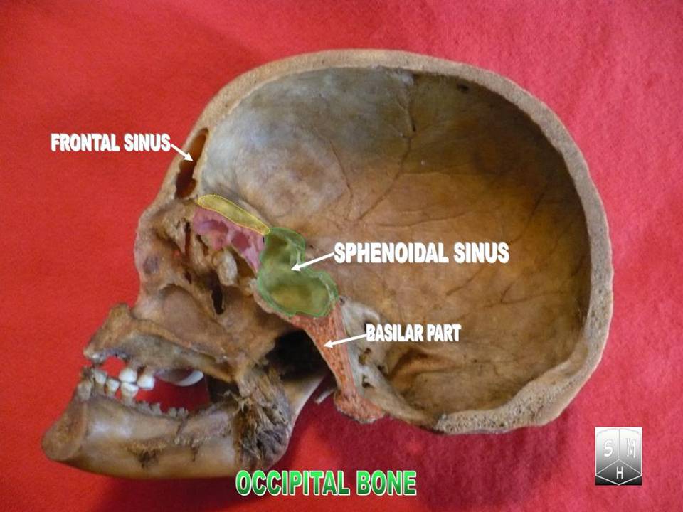


Anatomical parts and landmarks
The occipital bone can be divided into several regions: the squamous (posterior) part, the basilar (clival) part that extends toward the sphenoid, and paired lateral parts that contain the occipital condyles. Important surface landmarks include the external occipital protuberance and the superior and inferior nuchal lines, which serve as attachment sites for neck muscles. The hypoglossal canals traverse the bone near the condyles and transmit the hypoglossal nerves.
Development and articulations
During growth the occipital bone forms from multiple ossification centers that fuse over childhood and into early adulthood. It articulates with the two parietal bones, the two temporal bones and the sphenoid bone above and in front, and with the first cervical vertebra (atlas) via the occipital condyles below.
Clinical relevance
Because it surrounds the foramen magnum, the occipital bone is central in conditions that affect the craniovertebral junction. Trauma can produce basilar skull fractures; congenital or acquired crowding at the foramen magnum may contribute to Chiari malformations. Its muscle attachments make it a focus in tension-type or occipital neuralgia, and surgeons use occipital landmarks for posterior cranial approaches and decompression procedures.
- Protects posterior brain and cerebellum
- Provides muscle attachments for head and neck movement
- Contains the foramen magnum for neural and vascular passage
- Serves as an important surgical and radiographic landmark
Related articles
Author
AlegsaOnline.com Occipital bone — anatomy, function, and clinical relevance Leandro Alegsa
URL: https://en.alegsaonline.com/art/71819