Green fluorescent protein
Green fluorescent protein (GFP) is a naturally fluorescent protein used as a genetically encoded marker in biological research. This entry summarizes its structure, history, variants, and major applications.
Overview
Green fluorescent protein (GFP) is a naturally occurring protein that emits bright green light when exposed to blue or violet illumination. GFP was originally isolated from the jellyfish Aequorea victoria and is widely used as a genetically encoded fluorescent tag to visualize cells and subcellular structures or to report on molecular processes in living organisms.
Image gallery
10 Images


Structure and mechanism
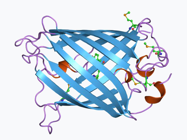
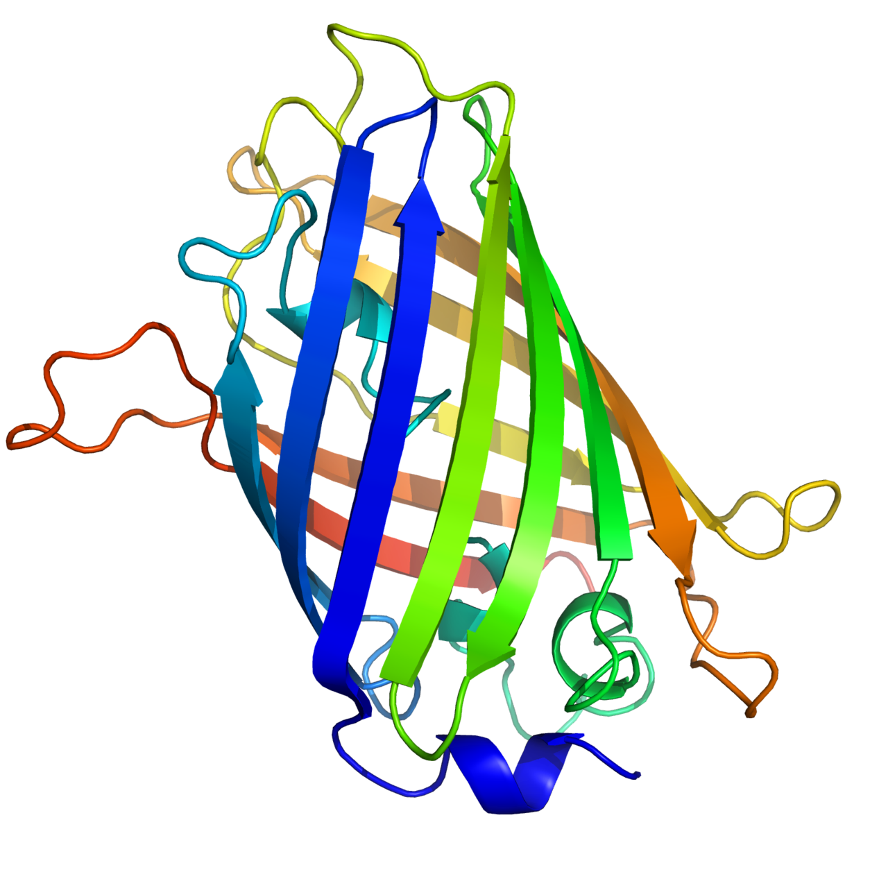
GFP's distinctive glow comes from a small internal chromophore formed by autocatalytic cyclization and oxidation of three amino acids in the protein chain. The protein adopts a sturdy beta-barrel fold that shields the chromophore and influences its spectral properties. When the chromophore absorbs light, it transitions to an excited state and then releases energy as green fluorescence—typically observed near 509 nm—making the molecule useful as a marker without needing added cofactors.
History and development
After its discovery and biochemical characterization, GFP was adapted for use in molecular biology by cloning its gene so it could be fused to other genes and expressed in bacteria, yeast, plants, and animals. Work on GFP and its engineered variants earned the 2008 Nobel Prize in Chemistry and stimulated a broad field of fluorescent-protein research and tool development.
Variants and engineering
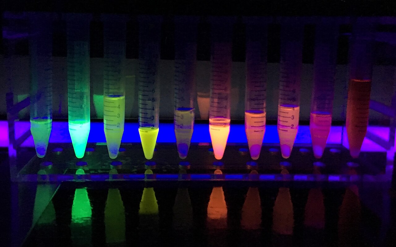
Researchers have altered GFP's sequence to change brightness, folding efficiency, color, and oligomeric state. Mutations produced enhanced forms such as EGFP and color-shifted derivatives: cyan (e.g., ECFP), yellow (YFP) and many others. Single amino-acid substitutions can shift excitation/emission wavelengths or improve photostability. In parallel, red fluorescent proteins derived from coral and other organisms (often termed RFP) expand the color palette for multicolor imaging; these are sometimes sourced from coral species.
Applications and examples
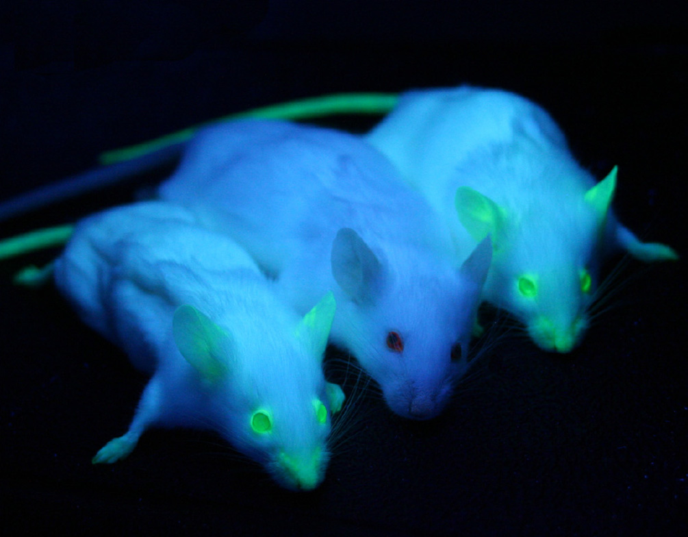
GFP and its relatives serve as fusion tags to follow protein localization, as reporters of gene expression, and as components of biosensors that reveal changes in ion concentration, enzyme activity, or protein interactions. Examples include calcium indicators based on GFP scaffolds and fluorescent reporters used in live-cell microscopy, developmental biology, and neuroscience. The genetic nature of GFP means it can be targeted to tissues or specific cell types, enabling time-lapse studies in living animals and cultured cells.
Practical considerations and notable facts
When using fluorescent proteins, researchers consider brightness, maturation time, pH sensitivity, photobleaching, and whether the protein behaves as a monomer (important when fusing to other proteins). The chromophore formation requires oxygen and proper folding, so cellular environment can affect fluorescence. Because changing or adding certain residues can alter color, scientists routinely engineer variants to create cyan, pink, and other hues for multiplexed imaging or improved performance; the family of fluorescent proteins remains a central imaging tool in modern biology.
For further reading on biochemical details and applications, see general resources on fluorescent proteins and practical guides to fluorescence microscopy. Many online and textbook sources provide protocols and comparisons of common GFP derivatives and their properties; consult specialized references for experimental planning and optimization.
More on molecular fluorescence · General fluorescence concepts
Related articles
Author
AlegsaOnline.com Green fluorescent protein Leandro Alegsa
URL: https://en.alegsaonline.com/art/40669