Grey matter (central nervous system tissue)
Grey matter is tissue in the brain and spinal cord made of neuronal cell bodies and glia; it mediates processing, integration and reflexes and contrasts with white matter rich in myelinated axons.
Overview
Grey matter is a primary tissue type of the central nervous system. It contains the cell bodies of neurons and supporting glial cells, together forming regions that perform local processing, integration of information and generation of outputs. In living brains the appearance of grey matter is pale grey with subtle pink or yellow tones from blood and metabolic pigments.
Image gallery
5 Images


Structure and components
Grey matter differs from white matter mainly by the presence of neuronal somata and short, often unmyelinated processes rather than long myelinated tracts. Key components include:
- Neuron cell bodies and dendrites, which receive and integrate synaptic input.
- Short-range axonal branches, frequently unmyelinated, that link nearby cells.
- Various types of glial cells that support metabolism, maintain ion balance and modulate synapses.
- Small blood vessels and capillaries that supply oxygen and nutrients.
The visual difference from white matter arises because myelin — formed by glia wrapping axons — is white, whereas grey matter has much less myelin; this contrast is driven by the presence or absence of extensively myelinated fibers and long axon tracts.
Function and examples
Grey matter houses the circuits responsible for perception, decision-making, voluntary movement planning and reflex control. Examples include the cerebral cortex, where layered grey matter supports high-level cognition, and subcortical nuclei (such as the basal ganglia) that regulate movement and reward. In the spinal cord, grey matter forms an inner H-shaped region that mediates reflexes and local integration.
Development, imaging and ageing
Grey matter develops through processes of neurogenesis, migration, synaptogenesis and later synaptic pruning. Its volume and composition change across life and can be measured noninvasively with imaging techniques; for instance, magnetic resonance imaging contrasts grey and white matter to study brain development and ageing. Small blood vessels and capillary networks contribute to the tissue's slightly colored appearance in vivo, observable at a microscopic level as reddish tones from capillary blood.
Clinical relevance and distinctions
Alterations of grey matter—loss, atrophy or focal damage—are central to many neurological conditions including stroke, traumatic injury and neurodegenerative diseases. Distinguishing grey from white matter helps clinicians localize lesions and understand symptoms, because damage to grey matter tends to disrupt local processing, while white matter damage impairs long-range communication.
Although terms and boundaries are simplified in broad descriptions, appreciating the cellular makeup and roles of grey matter is essential for studying brain function, development and disease.
Structure of the gray matter
In terms of developmental history, a distinction must be made between the substantia grisea centralis (central gray, cave gray) and the substantia grisea corticalis et intermedia (peripheral gray).
The peripheral gray has separated from the cavity system of the ventricles and the central gray found there. The peripheral gray is further subdivided into the cortical and the intermediate gray. It represents a peculiarity of the brain and is the seat of summary functions. No peripheral gray is found in the spinal cord. The intermediate grey of the brain (substantia grisea intermedia), which is to be distinguished from the substance of the same name in the spinal cord, forms the basal diencephalic nuclei (basal ganglia), nucleus hypothalamicus, substantia nigra, nucleus ruber, bridge nuclei, cerebellar nuclei, nucleus olivaris, etc., surrounded by white matter.
The cortical gray (substantia grisea corticalis) is characterized by layered structure or lamination. Here we can assume an organizing principle which, during the increase of the ganglion cell mass occurring in the course of development, does not allow the thickness of the cell masses to increase, but rather their areal expansion (surface enlargement). The excessive increase in surface expansion is counteracted by folding. This is how the outer formations of peculiarly winding gyri of the brain (substantia grisea corticalis), which are typical of the brain, are formed. But also in the intermediate gray, the folded cross-sectional images of nuclei such as the nucleus dentatus, or the nucleus olivaris are characteristic of this organizational principle. This planar spread of gray matter is found in the cerebrum and cerebellum, but also in the region of the superior tetrahedra. The advantage resulting from this principle is the better accessibility of the circuitry and therefore also of the retrieval, roughly comparable to the handiness of a chip card.
The central gray is considered within the brain as a nervous tissue connected to the ventricular system. The ventricular system has connection to the central canal in the area of the spinal cord. The entire cavity system arises from the clearing of the embryonic neural tube. The gray matter surrounding the central canal of the spinal cord is also called substantia grisea intermedia, see above. It has received this name because it connects the formations of the anterior horn and posterior horn, which are located on both sides of the spinal cord and also consist of gray matter, but it is not to be understood as substantia grisea intermedia in the sense of the developmental classification. Within the brain, the central gray is primarily the seat of the cranial nerve nuclei. The central cavernous gray represents the supreme center and the superior coordinating center for all vegetative functions. Such functions are heat and circulation regulation, digestion, excretion, sexual functions, etc.
Gallery
· 
Cross section of the spinal cord: white matter outside and grey matter inside.
· 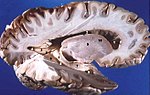
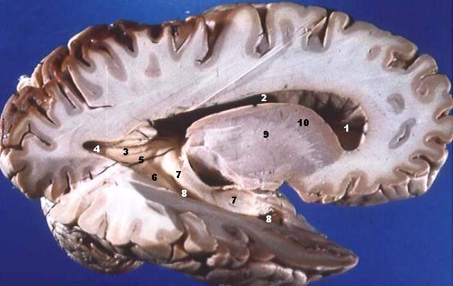
Sagittal section of the brain: gray matter outside and white matter inside.
Questions and answers
Q: What is grey matter?
A: Grey matter is a main component of the central nervous system, composed of neurons and glial cells.
Q: What is the difference between grey matter and white matter?
A: Grey matter is composed of neurons and glial cells, while white matter is composed of long-range myelinated axon tracts (nerve fibres) and glial cells.
Q: What is myelin?
A: Myelin is a substance that covers nerve fibres and gives white matter its whiteness.
Q: Why does grey matter have a different color from white matter?
A: The color difference arises mainly from the whiteness of myelin. In living tissue, grey matter actually has a very light grey color with yellowish or pinkish hues, which come from capillary blood vessels and neuronal cell bodies.
Q: What kind of cells make up grey matter?
A: Grey matter is composed of neurons (brain cells) and glial cells.
Q: What kind of cells make up white matter?
A: White matter is composed of long-range myelinated axon tracts (nerve fibres) and glial cells.
Q: What role does grey matter play in the central nervous system?
A: Grey matter is a main component of the central nervous system and plays an important role in processing information and controlling movement.
Related articles
Author
AlegsaOnline.com Grey matter (central nervous system tissue) Leandro Alegsa
URL: https://en.alegsaonline.com/art/40885