
Computed tomography (CT) scanner
A medical imaging device that uses X-rays to create cross-sectional images for diagnosis and treatment planning; covers technology, history, clinical uses, advantages, and limitations.
Overview
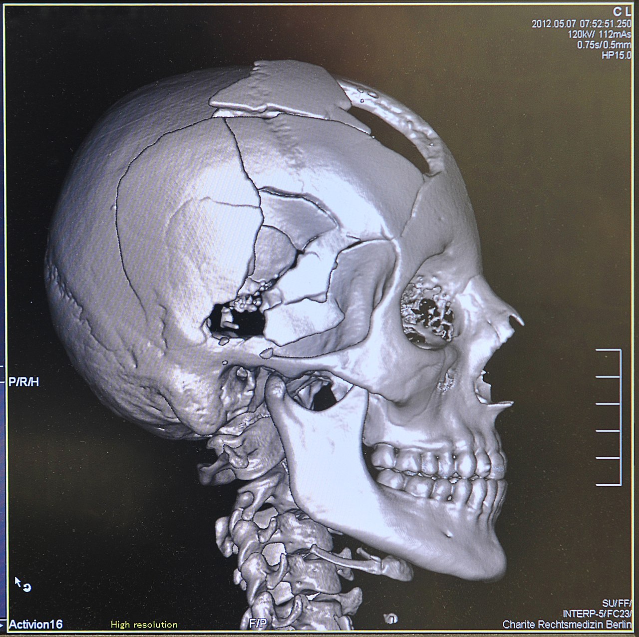
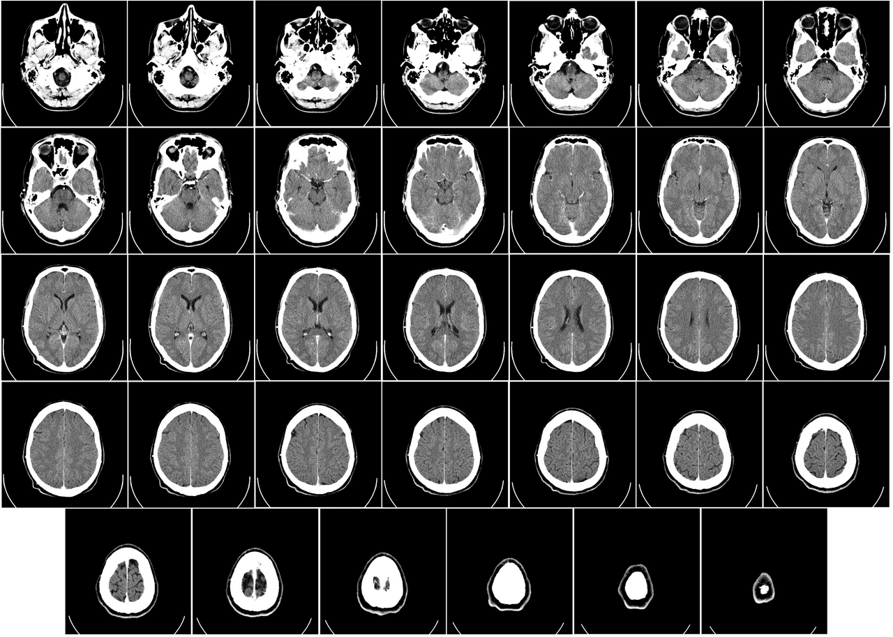
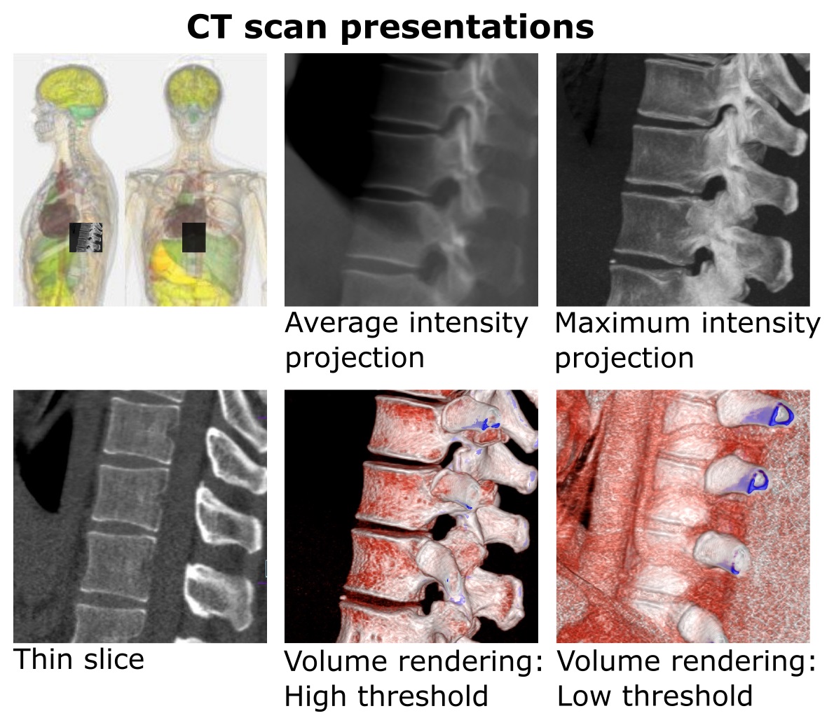
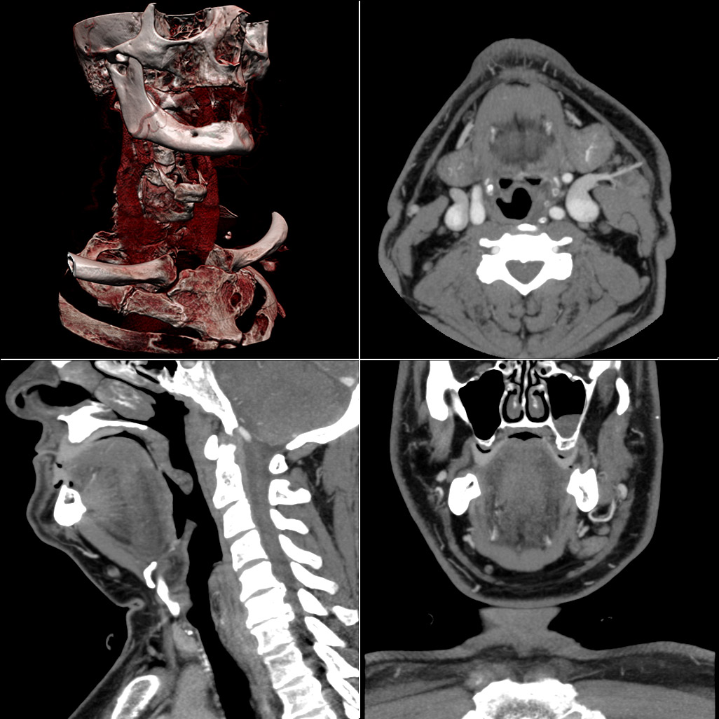
A computed tomography (CT) scanner is a medical imaging system that acquires detailed internal images by rotating an X-ray source and detectors around the patient. Modern CT produces a series of thin, cross-sectional images that can be reconstructed into three-dimensional views. A CT is typically used to visualize internal organs, bones, soft tissue and blood vessels for clinical assessment and planning.
Image gallery
10 Images



How it works
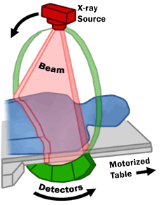
The scanner directs a narrow beam of radiation while detectors measure the transmitted intensity. Computer algorithms reconstruct many projection measurements into slice images. Typical components include:
- Gantry: the rotating ring that houses the source and detectors.
- Patient table: motorized couch that moves through the gantry.
- Detectors and X-ray tube: capture transmitted radiation and convert it to electrical signals.
- Reconstruction computer: turns raw data into cross-sectional images.
In routine descriptions the role of the radiation source is often referred to as X-ray generation, while the whole instrument is commonly called a machine. The resulting images are described as cross-sectional slices useful for clinical interpretation and diagnosis.
History and development
Computed tomography was developed in the late 1960s and early 1970s, with early pioneers combining X-ray measurements and computer reconstruction to produce sectional images. Subsequent innovations included helical (spiral) scanning, multi-detector arrays, faster rotation times and improved reconstruction methods, which increased speed, resolution and clinical utility.
Clinical uses and examples
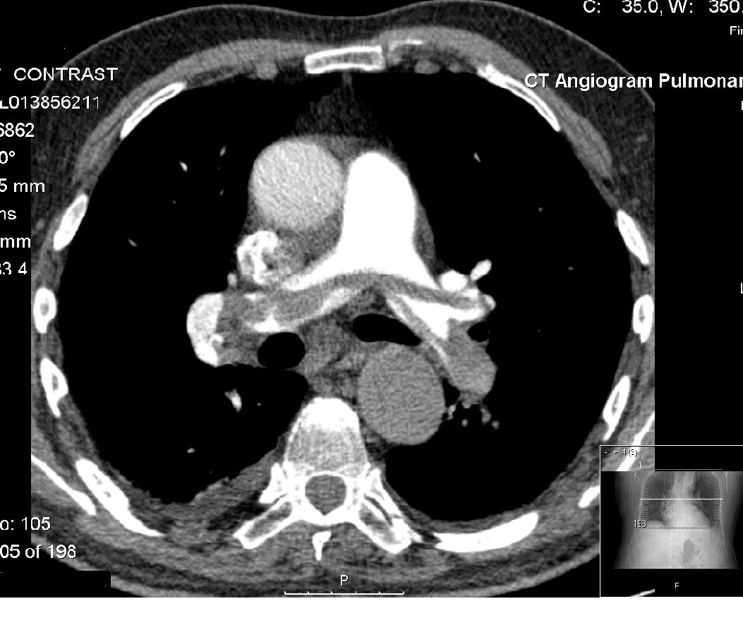
CT is widely used in emergency and elective care. Common indications include trauma evaluation, stroke assessment, chest and abdominal imaging, cancer staging, bone fractures and planning for surgery or radiotherapy. CT angiography visualizes blood vessels after injection of contrast agent. It is valued for speed, spatial detail and broad availability.
Advantages, limitations and notable facts
Advantages of CT include rapid acquisition, detailed bone and lung imaging, and high spatial resolution. Limitations include exposure to ionizing radiation, potential contrast reactions, and imaging artifacts from metal or patient motion. CT complements other modalities such as MRI and ultrasound; choice depends on clinical question, patient factors and availability.
Further reading
For introductions to technology, safety and clinical protocols consult manufacturer guidance, professional radiology societies and institutional resources. Technical reviews cover detector design, dose-reduction strategies and advanced applications such as volumetric perfusion, cardiac CT and spectral imaging.
Related articles
Author
AlegsaOnline.com Computed tomography (CT) scanner Leandro Alegsa
URL: https://en.alegsaonline.com/art/22300