Chimaera (genetics): organisms composed of genetically distinct cell lines
A chimaera is a single organism made from two or more genetically distinct cell populations. This article explains types, causes, detection, examples, and biological and medical significance.
Overview
In genetics, a chimaera (or chimera) is an individual whose body contains two or more populations of cells with different genotypes. Those distinct cell lines may derive from separate fertilized eggs that fused early in development, or from other processes that introduce genetically distinct cells into a host. The term covers a range of situations from visibly mosaic pigmentation to microscopic populations of foreign cells that have clinical or forensic importance. For a basic description of origins, see fertilized eggs and early embryo fusion.
Image gallery
6 Images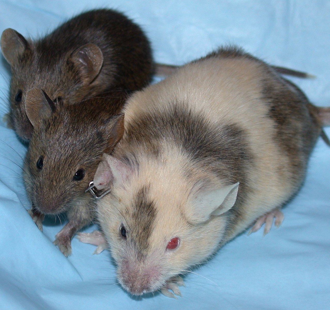


Key forms and how they arise
Chimaeras are commonly classified by origin and extent of mixing. Important categories include:
- Dispermic or tetragametic chimaeras — arise when two (or more) zygotes or early embryos merge; the resulting individual contains large cell patches from each original embryo and often shows mixed phenotypes such as differing skin patches or organ-level contributions. Further reading: distinct cell populations and phenotypes.
- Mosaicism — technically different from chimaerism when the genetically distinct cells arise by mutation or segregation events within a single zygote; mosaic individuals tend to show more limited or patterned variation. See a comparison at mosaic versus chimaera.
- Microchimerism — the presence of a small number of foreign cells in an individual, often clinically silent. Common sources are pregnancy (bidirectional cell trafficking between mother and fetus), transfusion, or transplantation; summaries are available at maternal–fetal cell exchange and transfusion-related or transplant-associated microchimerism.
- Epigenetic chimaeras — cases in which cell populations differ in gene expression regulation rather than DNA sequence; such patterns have been observed in laboratory mouse models and are under active study (epigenetic variation).
Characteristics and detection
Observable traits of chimaerism range from striking to cryptic. Visible signs can include asymmetric or patchy skin pigmentation, mismatched eye color, or differences in blood group across tissues. Many chimaeras are only detected by genetic testing of multiple tissues or by forensic profiling when unexplained genotype mixtures appear. Clinically, microchimerism can be identified by sensitive molecular assays that detect low-frequency alleles.
Biological and medical importance
Chimaerism has practical consequences in several fields. In medicine it can complicate organ matching, diagnosis of genetic disease, and interpretation of parentage or forensic tests. In developmental biology and regenerative medicine, deliberate chimaeras are powerful experimental tools for tracing cell fates and testing stem cell potential. Natural examples, such as freemartinism in cattle where twin embryos exchange cells and hormones, illustrate both ecological and agricultural relevance.
History, research, and ethical notes
Recognition of chimaerism dates to anatomical and cytogenetic observations in the 20th century; modern molecular methods greatly increased detection sensitivity. Experimental creation of chimaeras has advanced knowledge of development but also raises ethical considerations when applied to human embryos or interspecies combinations. Ongoing research explores how microchimeric cells influence immunity, tissue repair, and autoimmunity, and how epigenetic heterogeneity affects phenotype without sequence changes.
For broader context and practical guides, consult introductory resources on embryology and molecular genetics as well as specialized reviews on microchimerism and mosaicism. More links: early embryology, mosaicism overview, phenotypic consequences, transfusion impacts, transplant impacts, pregnancy and cell trafficking, epigenetics research.
Questions and answers
Q: What is a chimaera in biology?
A: A chimaera is a single organism made of two genetically distinct cells, usually an animal.
Q: How do chimeras originate?
A: Chimeras can originate from two separate fertilized eggs or zygotes that fused together, or from the same zygote, in which case the organism is called a mosaic.
Q: What are dispermic chimeras?
A: Dispermic chimeras are chimeras that originate from distinct fertilized eggs that fused together.
Q: How are dispermic chimeras formed?
A: Dispermic chimeras are formed from at least two fertilized eggs or early embryos fused together.
Q: How do the different populations of cells in a dispermic chimera behave?
A: Each population of cells in a dispermic chimera keeps its own phenotype, and the resulting organism is a mixture of the two phenotypes.
Q: What is microchimerism?
A: Microchimerism is a form of chimerism in which a host harbors a small set of genetically distinct cells, often due to blood transfusion, transplant or pregnancy.
Q: What are epigenetic chimeras?
A: Epigenetic chimeras are chimeras that have been recently discovered in mouse models, in which the genetic information of some cells is modified by the environment without altering the DNA sequence itself.
Related articles
Author
AlegsaOnline.com Chimaera (genetics): organisms composed of genetically distinct cell lines Leandro Alegsa
URL: https://en.alegsaonline.com/art/19713
Sources
- pnas.org : "Germ-line chimerism and paternal care in marmosets (Callithrix kuhlii)"
- doi.org : 10.1073/pnas.0607426104
- worldcat.org : 0027-8424
- ncbi.nlm.nih.gov : 1851065
- pubmed.ncbi.nlm.nih.gov : 17389380
- nytimes.com : "In the marmoset family, things really do appear to Be all relative"
- newscientist.com : "Marmosets may carry their sibling's sex cells"