
Wound healing: processes, stages and clinical considerations
Comprehensive overview of wound healing: phases (hemostasis, inflammation, proliferation, remodeling), key cells and molecules, factors that help or hinder repair, complications and basic care principles.
Overview
Wound healing is the biological process by which the body repairs damaged tissue after injury. Intact skin consists of an outer epidermis and an underlying dermis that together form a barrier; damage to these layers triggers a coordinated cascade of events to restore tissue continuity. The earliest responses include blood clot formation and immune activation, followed by tissue growth and long-term remodeling. The epidermis can re-form by migration of surface cells (epidermis) while deeper structures are rebuilt by connective tissues.
Image gallery
8 Images

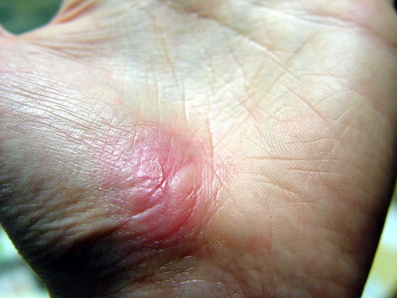
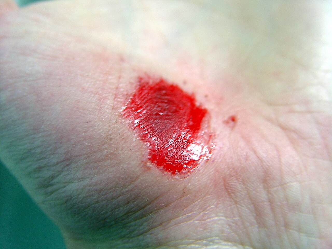




Phases of healing
Healing is commonly described in four overlapping phases. These stages are predictable but not strictly sequential; they overlap and vary with wound type and patient condition:
- Hemostasis: immediate vascular constriction and platelet-driven clot formation to stop bleeding and form a provisional matrix (hemostasis).
- Inflammation: neutrophils and macrophages clear debris and microbes; inflammation sets the biochemical environment for repair (inflammation).
- Proliferation: fibroblasts produce extracellular matrix, endothelial cells form new capillaries (angiogenesis), and epithelial cells migrate to cover the defect.
- Remodeling (maturation): collagen is reorganized and strengthened; scar tissue forms and gradually changes over weeks to months.
Cellular and molecular components
Multiple cell types and signaling molecules coordinate healing. Platelets release growth factors that attract immune cells. Neutrophils provide early microbial control, while macrophages switch from pro-inflammatory to pro-repair roles and stimulate fibroblasts. Fibroblasts synthesize collagen and other matrix proteins; keratinocytes re-epithelialize the surface. Key molecular players include cytokines, growth factors (e.g., PDGF, TGF-β), matrix metalloproteinases and their inhibitors, and different collagen types that determine scar architecture.
Factors influencing repair
Many systemic and local factors affect the speed and quality of healing. Favorable conditions include good blood supply, adequate oxygenation, balanced moisture, and proper nutrition. Impairing factors include infection, poor perfusion (for example from vascular disease or smoking), diabetes, malnutrition, advanced age, certain medications (like corticosteroids), and continued mechanical stress on the wound. Wounds may heal by primary intention (edges approximated) or secondary intention (large gaps filled in by granulation tissue).
Complications and clinical considerations
Normal repair may be complicated by infection, delayed healing, chronic non-healing wounds (such as some pressure ulcers or diabetic ulcers), excessive scar formation (hypertrophic scars or keloids), and contractures that limit function. Basic clinical care principles are control of bleeding, removal of contaminants and devitalized tissue (debridement), prevention or treatment of infection, and use of dressings that maintain an appropriate moisture balance. Advanced treatments include negative-pressure wound therapy, topical growth factors, skin grafts and, when indicated, surgical closure.
History, distinctions and notable facts
Concepts of wound care date to ancient medicine, but understanding improved markedly after the germ theory and the discovery of antisepsis and suturing techniques. Modern research distinguishes true regeneration—restoring tissue identical to the original—from repair, where scar tissue replaces lost structures. Some animals regenerate whole structures with little scarring, whereas humans typically repair with a scar that continues to remodel long after surface closure. After healing finishes, a visible scar commonly remains (scar), and the general process described here does not apply to animals protected by a rigid external covering such as an exoskeleton (exoskeleton).
For clinicians and caregivers, recognizing the phase of healing, identifying modifiable impediments, and applying appropriate local care are central to improving outcomes. Further information on anatomy of the epidermis and deeper skin layers, molecular mediators, and advanced therapies can be found via specialist resources (epidermis basics, hemostasis overview, inflammatory response, scar management, comparative biology).
Related articles
Author
AlegsaOnline.com Wound healing: processes, stages and clinical considerations Leandro Alegsa
URL: https://en.alegsaonline.com/art/109159
Sources
- books.google.com : Wound healing · web.archive.org
- ncbi.nlm.nih.gov : "The role of nuclear hormone receptors in cutaneous wound repair"
- doi.org : 10.1002/cbf.3086
- pubmed.ncbi.nlm.nih.gov : 25529612
- doi.org : 10.1016/S0002-9610(98)00183-4
- pubmed.ncbi.nlm.nih.gov : 9777970