
Ulna: anatomy, function, and clinical importance
The ulna is the medial bone of the forearm. This article explains its parts, articulation with the radius and humerus, role in movement, variation, and common injuries.
Overview
The ulna is one of the two long bones that form the lower forearm in many vertebrates. In humans it lies on the side of the little finger and works together with the radius. The ulna contributes to the elbow joint above and the wrist and hand below, transmitting forces from the hand to the upper limb. It is present across a wide range of tetrapod species, where its shape and proportions reflect different modes of locomotion and manipulation (tetrapods).
Image gallery
10 Images




Anatomical features
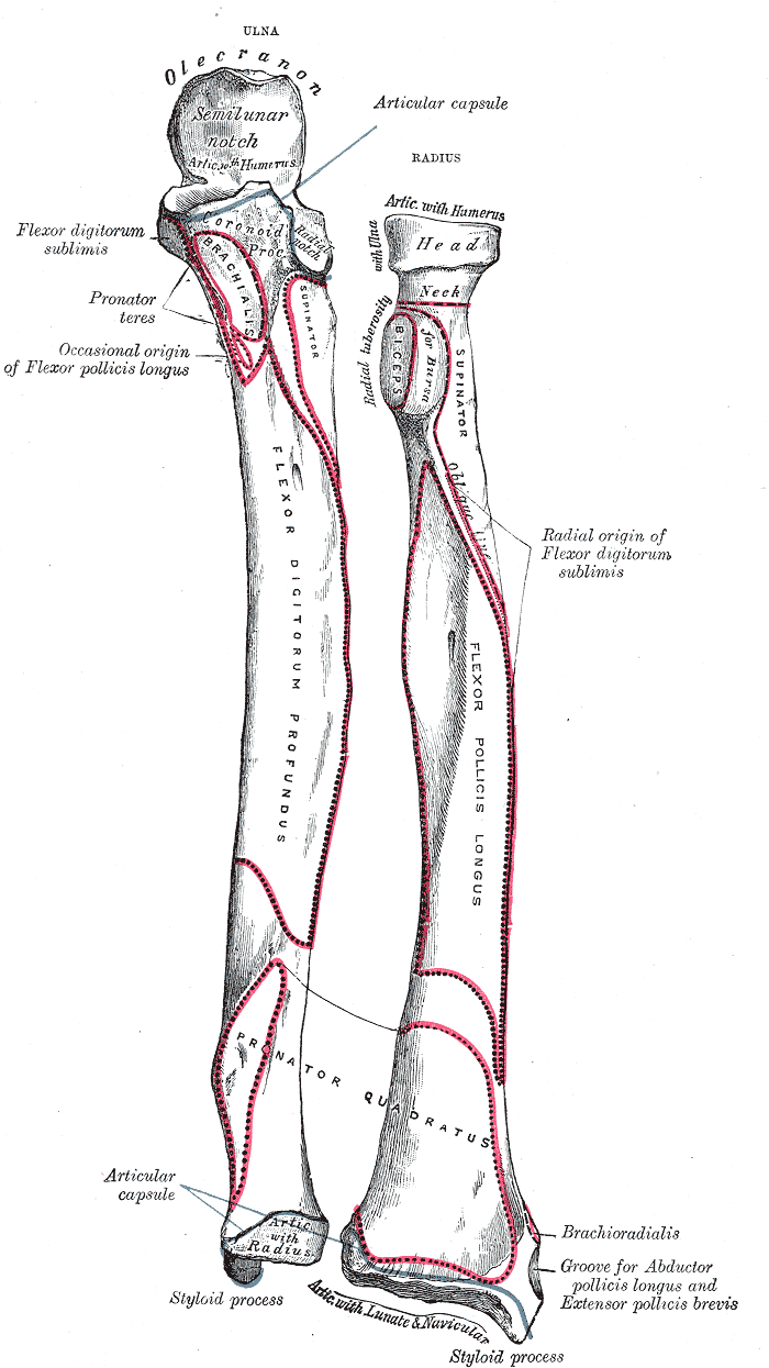
The ulna can be described in three regions: proximal end, shaft, and distal end. The proximal end includes the olecranon — a prominent posterior projection that forms the bony tip of the elbow — and the trochlear notch, which articulates with the humerus. The shaft is slightly triangular in cross-section and gives attachment to muscles and interosseous membrane. The distal end is smaller and participates in the distal radioulnar joint, allowing rotation with the radius.
- Proximal: olecranon, coronoid process, trochlear notch (articulates with humerus).
- Shaft: surfaces for muscle attachment and interosseous membrane (connects to the other forearm bone).
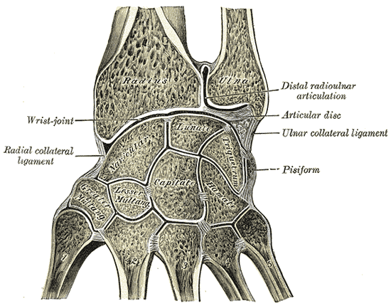
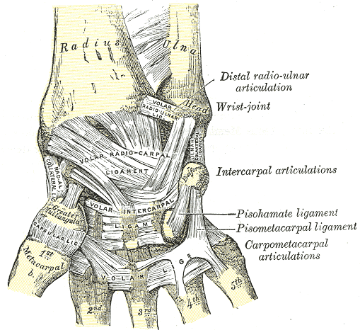
- Distal: head and styloid process, forms part of the wrist and the joint with the radius.
Function and movement
Functionally, the ulna stabilizes the forearm and, through its articulations, allows flexion and extension at the elbow and rotation (pronation and supination) of the forearm when the radius pivots around it. Muscles such as the triceps brachii insert on the olecranon to extend the elbow, while many forearm flexors and extensors attach along the shaft.
Clinical significance and variation
Injury to the ulna includes fractures (isolated or combined with the radius), dislocations affecting the proximal or distal radioulnar joints, and conditions such as osteoarthritis. An example of a combined injury is the Monteggia fracture-dislocation, which involves the proximal ulna and the radial head. Anatomical variation occurs between species and among individuals; in some animals the ulna is reduced or fused for specialized limb function. For additional anatomical references see resources on forearm anatomy (lower forearm) and comparative skeletal anatomy (elbow and humerus relations).
Understanding the ulna's form and connections is important in medicine, orthopedics, anthropology and comparative anatomy, as its shape reflects both mechanical demands and evolutionary history.
Related articles
Author
AlegsaOnline.com Ulna: anatomy, function, and clinical importance Leandro Alegsa
URL: https://en.alegsaonline.com/art/102672