Medical ultrasonography: principles, applications, and clinical use
Overview of medical ultrasonography covering how ultrasound imaging works, common clinical applications (obstetrics, cardiac, emergency), key techniques, history, safety, advantages and limitations.
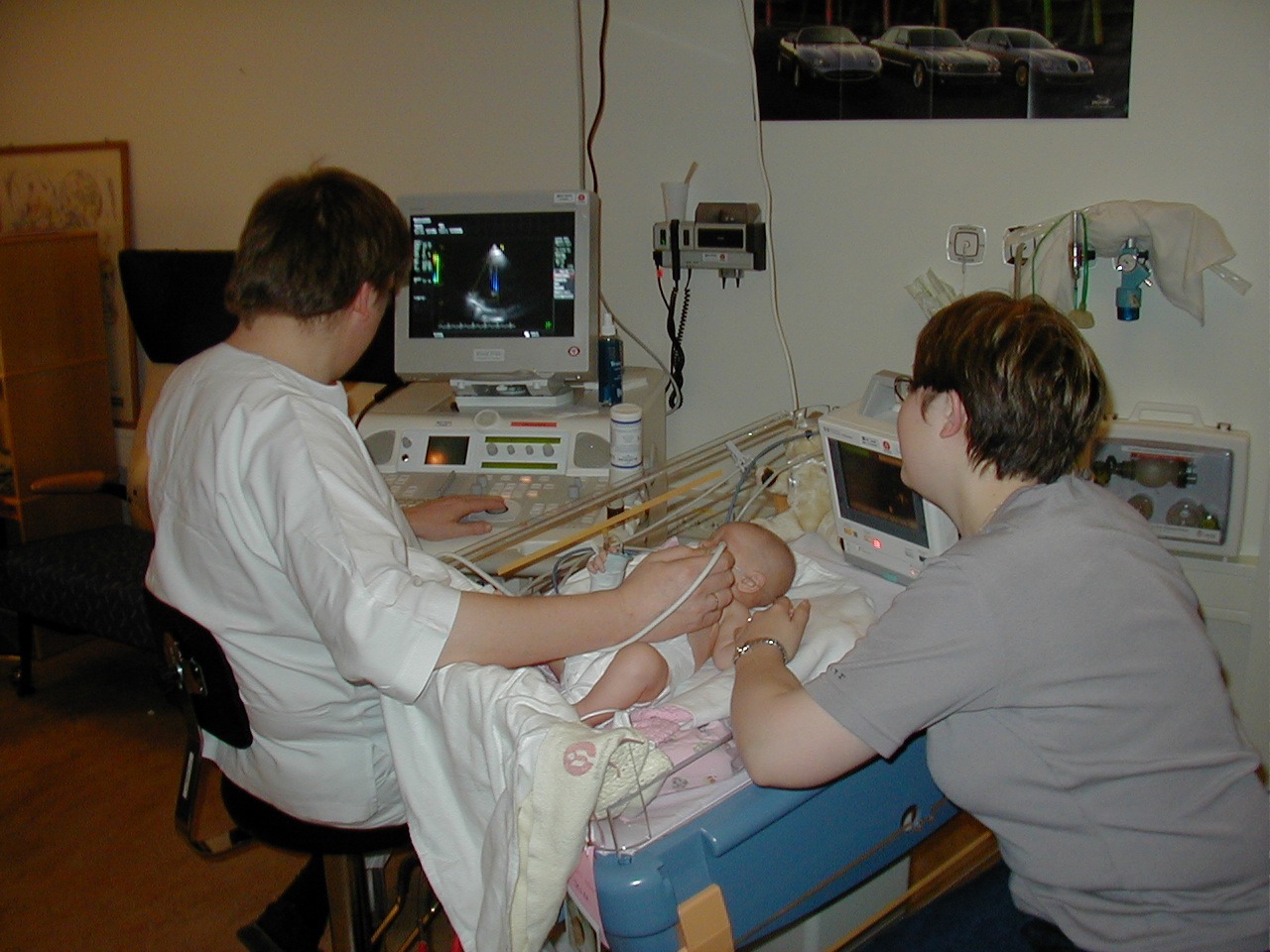
Medical ultrasonography, often called diagnostic ultrasound or sonography, uses high-frequency sound waves to produce images of structures inside the body. A handheld probe (transducer) sends pulses of sound which bounce off tissues and return echoes that are converted into realtime images. Because it relies on non-ionizing sound rather than X-rays, ultrasonography is widely used for imaging organs, soft tissues and blood flow and for guiding minimally invasive procedures.
Image gallery
10 Images


How it works and main components
The core elements of an ultrasound system are the transducer, the beamforming and signal-processing electronics, and the display. The transducer contains piezoelectric crystals that generate and detect sound waves at frequencies typically between 1 and 20 MHz. Differences in acoustic impedance between tissues produce echoes; the system measures echo timing and amplitude to construct two-dimensional (B-mode) or specialized images. Doppler techniques analyze frequency shifts to show blood flow velocity and direction, and color Doppler overlays flow information onto anatomic images.
Common clinical applications
- Obstetrics and prenatal care: fetal growth, placental location and basic anomaly screening. See pregnancy imaging for routine prenatal assessments.
- Abdominal imaging: liver, gallbladder, kidneys, spleen and biliary tree evaluation.
- Cardiac ultrasound (echocardiography): structure and function of heart chambers and valves.
- Vascular studies: evaluation of arteries and veins, stenosis and thrombosis using Doppler.
- Musculoskeletal imaging: assessment of muscles, tendons and soft tissue masses.
- Emergency and point-of-care use: rapid bedside exams in emergency medicine, including the Focused Assessment with Sonography for Trauma (trauma or FAST) to detect free fluid in body cavities.
Ultrasonography also plays a central role in procedural guidance—helping clinicians place needles for biopsies, drainage or vascular access with improved accuracy and safety. Portable and handheld devices have expanded access to bedside imaging and non-radiology environments.
History and development
Medical ultrasound grew out of acoustic and sonar research in the first half of the 20th century and entered clinical practice in the mid-1900s. Early pioneers adapted ultrasound for organ imaging and obstetrics; subsequent advances in electronics, transducer design and digital signal processing enabled the detailed, real-time images commonly used today. Modern developments include three-dimensional and four-dimensional imaging, high-frequency probes for superficial structures, and contrast-enhanced ultrasound for selective vascular or lesion characterization.
Advantages, limitations and safety
Key advantages of ultrasonography are its non-ionizing nature, relative affordability, portability and ability to produce dynamic, realtime images. It is well suited for evaluating soft tissues and blood flow, and for guiding interventions. Limitations include operator dependence, reduced image quality in obese patients or when gas obscures the acoustic window, and lower sensitivity for air-filled or calcified structures. Although generally regarded as safe, prudent use follows established guidelines regarding exposure and thermal/mechanical indices.
Techniques and notable facts
Specialized modes include Doppler, color-flow mapping, elastography to estimate tissue stiffness, and contrast-enhanced studies using microbubble agents for improved vascular imaging. Training and credentialing vary by specialty; accurate interpretation requires knowledge of anatomy, sonographic artifacts and clinical context. For rapid assessment of patients in shock, focused protocols can provide lifesaving information. Educational resources and clinical guidelines are available through professional societies and institutional programs—consult authoritative sources for local practice standards and training pathways (see links and references: ultrasound, tendons).
Related articles
Author
AlegsaOnline.com Medical ultrasonography: principles, applications, and clinical use Leandro Alegsa
URL: https://en.alegsaonline.com/art/63421