
Melanocyte: pigment-producing skin cells and their roles
Melanocytes are specialized cells that synthesize melanin, contributing to skin, hair and eye color and protecting against UV damage. This article covers structure, development, regulation, disorders and clinical relevance.
Overview
Melanocytes are specialized cells that manufacture the pigment melanin. They are most prominent in the epidermis of the skin, in hair follicles, and in the iris of the eye. Melanin itself is the pigment responsible for much of the visible variation in skin and hair color; it also contributes to the shading of internal tissues. For a concise definition of melanin see melanin and for the general concept of pigment see pigment.
Image gallery
3 Images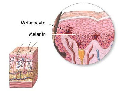

Structure and function
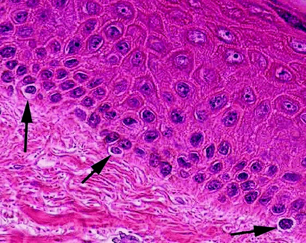
Melanocytes have a distinctive shape with branching dendrites that contact neighboring keratinocytes. Within each cell, pigment is synthesized and stored in membrane-bound organelles called melanosomes. The enzyme tyrosinase catalyzes early steps of melanin production; the balance between eumelanin (brown/black) and pheomelanin (red/yellow) helps determine visible color. Melanosomes are transferred from melanocytes to keratinocytes, distributing pigment across the epidermis.
Development and distribution
In embryonic development melanocytes arise from neural crest cells and migrate to the skin, hair follicles and eyes. Their numerical density is similar across human populations; differences in skin tone are largely due to the amount, type and packaging of melanin rather than simply cell count. Melanocytes can also be found in mucous membranes and other internal sites in lower numbers.
Regulation and photoprotection
Melanin absorbs ultraviolet (UV) radiation from the Sun, reducing photochemical damage to cellular components such as DNA. Exposure to UV stimulates melanin synthesis and distribution, producing tanning. Hormonal signals modulate pigment production: melanocyte-stimulating hormones (MSH) from the pituitary gland bind melanocortin receptors (notably MC1R) on melanocytes, increasing melanin synthesis. MSH-related pathways also interact with central pathways that influence behavior, including effects on brain circuits linked to appetite and sexual arousal, illustrating biochemical overlap between pigmentation and broader physiology.
Clinical importance and common disorders
Melanocyte function is central to a range of medical conditions. Reduced or absent melanin synthesis causes forms of albinism, often due to tyrosinase defects, leading to very light skin and increased photosensitivity. Autoimmune destruction of melanocytes produces vitiligo, characterized by well-demarcated depigmented patches. Overproliferation of melanocytes gives rise to melanoma, the most serious form of skin cancer; UV exposure is a major environmental risk factor for DNA damage and malignant transformation. More benign changes include lentigines and nevi (moles), which reflect localized variations in melanocyte activity or number.
Research, cosmetic and societal aspects
Melanocyte biology is an active research area across dermatology, oncology and genetics. Studies examine pigmentation genetics, pathways of melanosome transfer, and therapies to restore pigment or prevent melanoma. Clinically and cosmetically, melanin influences sun protection advice, pigmentation treatments, and social perceptions of appearance. For further reading on related topics see background resources on hair, the epidermis, and clinical information about skin cancer.
- Key features: melanosomes, tyrosinase, dendritic transfer
- Functions: determine color, absorb UV, modulate oxidative stress
- Clinical notes: albinism, vitiligo, melanoma — prevention emphasizes UV protection
Questions and answers
Q: What are melanocytes?
A: Melanocytes are the cells that produce melanin.
Q: Where are melanocytes most obvious?
A: Melanocytes are most obvious in the skin, hair, and eyes.
Q: Where are melanocytes located in the skin?
A: Melanocytes are located in the bottom layer of the skin's epidermis.
Q: What is melanin responsible for?
A: Melanin is mostly responsible for skin and hair color.
Q: What does melanin do to protect the skin?
A: Melanin protects the skin against damage by UV radiation from the Sun.
Q: What causes skin cancer?
A: UV radiation causes damage to cell DNA, which makes skin cancer more likely.
Q: What stimulates melanocytes to produce more melanin?
A: Melanocyte-stimulating hormones (MSH) from the pituitary gland stimulate melanocytes to produce more melanin.
Related articles
Author
AlegsaOnline.com Melanocyte: pigment-producing skin cells and their roles Leandro Alegsa
URL: https://en.alegsaonline.com/art/63627
Sources
- en.oxforddictionaries.com : "Melanocyte"
- merriam-webster.com : "Melanocyte"
- doi.org : 10.1016/0361-9230(83)90218-6
- pubmed.ncbi.nlm.nih.gov : 6616275