Kidney stone disease (urolithiasis): causes, symptoms, diagnosis and treatment
Kidney stone disease (urolithiasis) is formation of solid mineral deposits in the urinary tract. This article explains types, symptoms, diagnosis, treatments, prevention and notable facts.
Overview
Kidney stone disease, or urolithiasis, is the formation of hard mineral and salt deposits within the urinary system. Most stones begin in the kidney (kidney) and can travel down the urinary tract, sometimes passing in urine (urination). Stone size varies from tiny grains to objects several millimetres across. Small stones often pass unnoticed; larger ones can obstruct urine flow and cause intense symptoms.
Image gallery
10 Images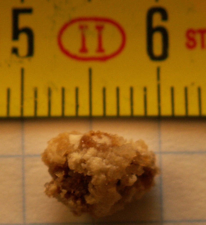


Causes, composition and risk factors
Stones form when urine contains high concentrations of stone-forming substances such as calcium, oxalate, uric acid, or cystine, or when protective factors are low. Common stone types include calcium oxalate, uric acid, struvite (often infection-related), and cystine (genetic). Risk factors include dehydration, certain diets, metabolic conditions, family history, obesity, and recurrent urinary infections.
Symptoms and complications
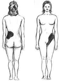
When a stone lodges in a narrow part of the urinary tract, particularly the ureter (ureter), it often causes sudden, severe pain in the flank, lower back or abdomen (pain) and may produce blood in the urine, nausea, and vomiting (vomiting). Complications include urinary obstruction, recurrent infections, reduced kidney function, and chronic pain. About half of people who form a stone will experience a recurrence within ten years.
Diagnosis
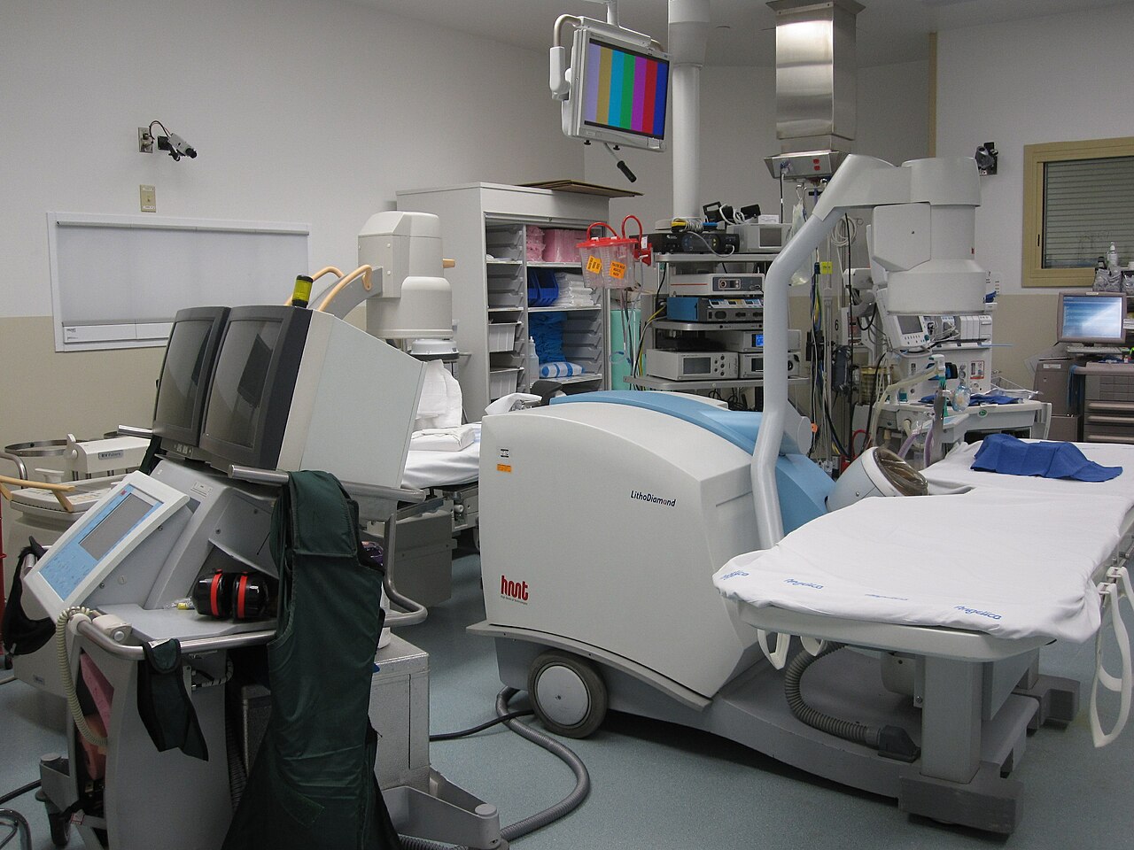
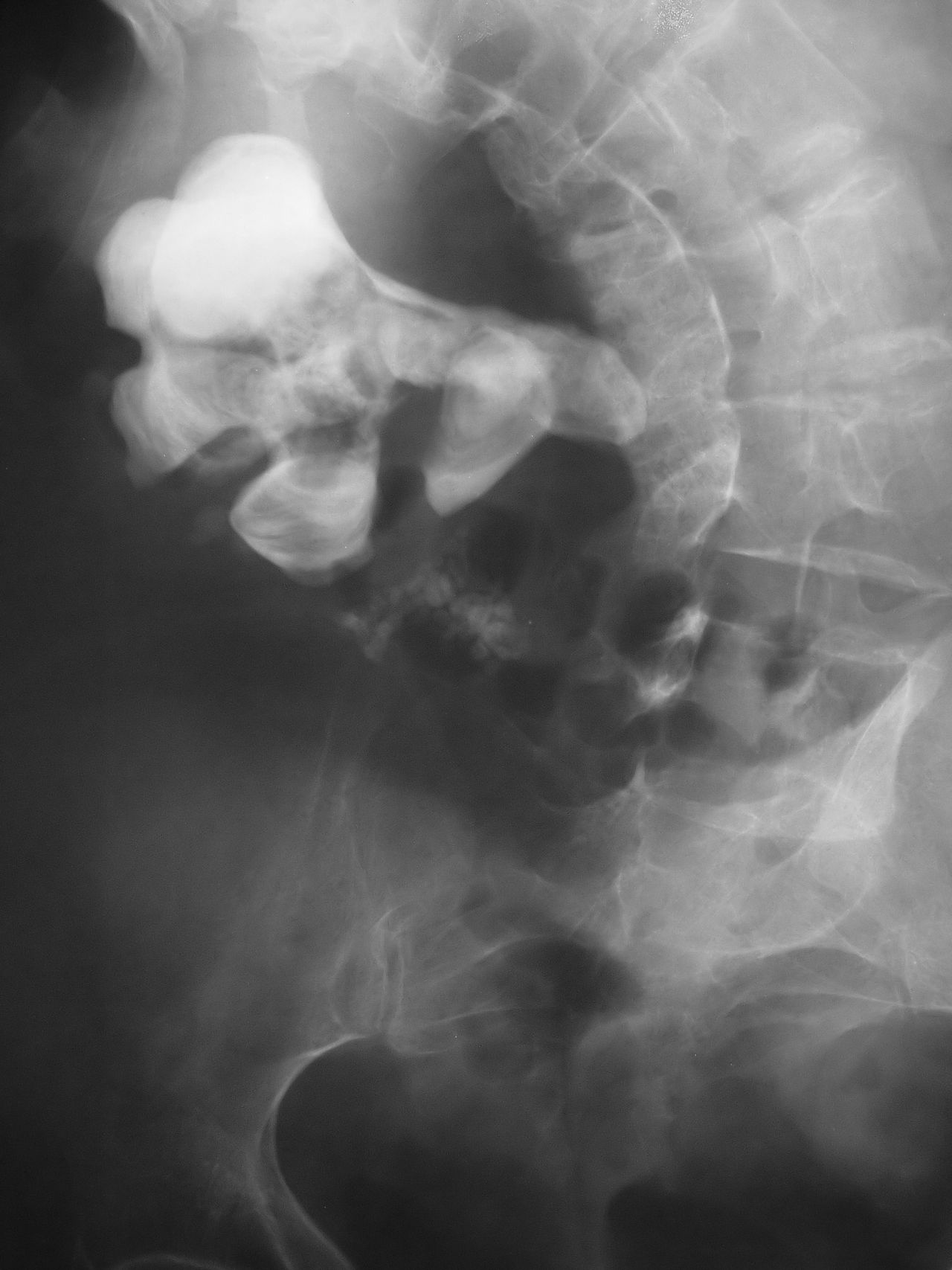
Diagnosis typically combines clinical history, urinalysis and imaging. Non-contrast CT scanning is highly sensitive for detecting stones; ultrasound is a radiation-free alternative often used in pregnancy or initial screening. Urine testing can identify crystals, infection, and metabolic contributors. Blood tests check kidney function and levels of minerals.
Treatment and prevention
Management depends on stone size, location and symptoms. Small stones may pass with increased oral fluids and pain control. Medications can help relax the ureter. Larger or obstructing stones may require extracorporeal shock wave lithotripsy, ureteroscopy with laser fragmentation, or percutaneous removal. Long-term prevention focuses on raising fluid intake, dietary adjustments, and targeted medications when metabolic abnormalities are identified.
History and notable facts
Kidney stones have been recognised since antiquity and clinical approaches have evolved from open surgery to minimally invasive and noninvasive interventions. Prompt evaluation of severe pain and suspected obstruction reduces the risk of complications. For further reading and patient resources see links provided above and consult medical professionals for personalized advice.
Related articles
Author
AlegsaOnline.com Kidney stone disease (urolithiasis): causes, symptoms, diagnosis and treatment Leandro Alegsa
URL: https://en.alegsaonline.com/art/53331
Sources
- niddk.nih.gov : "Kidney Stones in Adults"
- books.google.com : Clinical Management of Urolithiasis · web.archive.org
- bmj.com : "Management of kidney stones" · web.archive.org
- doi.org : 10.1136/bmj.39113.480185.80
- ncbi.nlm.nih.gov : 1808123
- pubmed.ncbi.nlm.nih.gov : 17332586