Intercostal muscles: anatomy, function, and clinical significance
Intercostal muscles are layers of skeletal muscle between the ribs that support breathing, chest-wall stability and movement. This article covers anatomy, action, innervation and clinical relevance.
The intercostal muscles are bands of skeletal muscle that occupy the spaces between adjacent ribs. They form part of the chest wall and act together with the diaphragm and other thoracic muscles to change thoracic volume during breathing. By altering the position of the ribs and the stiffness of the rib cage, the intercostals contribute to ventilation, protection of internal organs, and mechanical stability of the torso.
Image gallery
5 Images

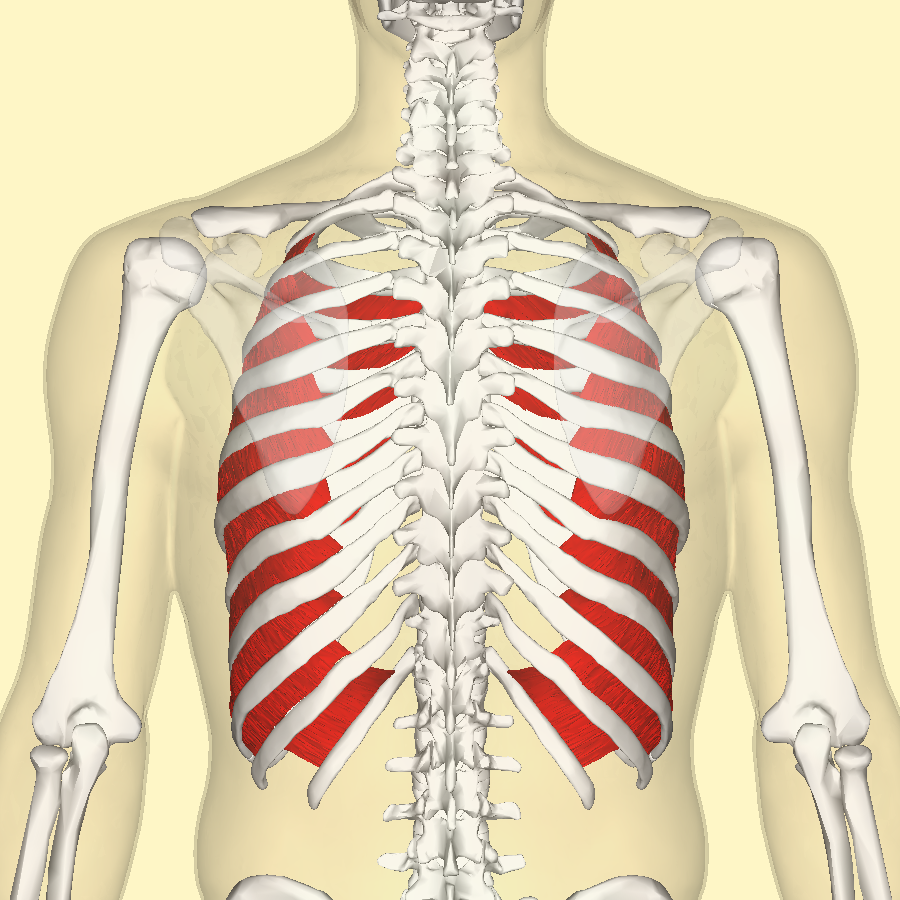


Anatomy and layers
Intercostal muscles are usually described in three layers, each with distinct fiber directions and attachments. They extend from the tubercles of the ribs posteriorly to the costal cartilages anteriorly and fill the intercostal spaces:
- External intercostal muscles — superficial layer; fibers run obliquely downwards and forwards (often remembered as "hands in pockets"). They primarily assist with elevating the ribs during quiet and forced inspiration.
- Internal intercostal muscles — middle layer; fibers run at roughly right angles to the externals, downwards and backwards. They are active in forced expiration, drawing the ribs downward.
- Innermost intercostal muscles — deepest and discontinuous; work with the internal layer and lie adjacent to the neurovascular bundle embedded in the costal groove.
Function and mechanics
During inhalation the external intercostals contract to lift and widen the rib cage, increasing thoracic volume and aiding inflow of air. Exhalation at rest is largely passive, but internal and innermost intercostals assist in forced expiration by depressing the ribs. The intercostals also stabilize the thorax during movements of the upper limbs and trunk, and prevent paradoxical motion of the chest wall. These muscles act in concert with the diaphragm, accessory respiratory muscles, and pleural mechanics to maintain effective ventilation.
Innervation, blood supply and clinical notes
Each intercostal muscle is supplied by the corresponding intercostal nerve, a ventral ramus of a thoracic spinal nerve, and receives arterial blood from intercostal arteries. Clinically, intercostal muscles and their nerves are important in procedures such as intercostal nerve blocks and thoracostomy; trauma or surgical injury can cause pain, impaired breathing, or intercostal neuralgia. Signs such as intercostal retraction are observed in respiratory distress. Strain or spasm of these muscles may produce localized chest pain that can mimic other conditions.
In anatomy and surgery the arrangement of the neurovascular bundle—typically running between the internal and innermost layers—is an important landmark. Care is taken to avoid this bundle when inserting needles or chest drains. Variations in muscle thickness and activity occur with age, posture, fitness, and pulmonary disease.
Origins, terminology and broader importance
The term "intercostal" derives from Latin roots meaning "between the ribs." Anatomical description of these muscles dates back centuries and they remain a fundamental subject in studies of respiration. Beyond respiration, intercostal muscles influence posture, coughing mechanics, and the transmission of forces through the thorax. For further anatomical images and functional summaries see linked resources: rib and thorax overview, respiratory mechanics, and clinical guidance on nerve blocks and interventions at procedural notes.
Related articles
Author
AlegsaOnline.com Intercostal muscles: anatomy, function, and clinical significance Leandro Alegsa
URL: https://en.alegsaonline.com/art/47580
Sources
- commons.wikimedia.org : Intercostal muscle
- healthline.com : "BodyMaps; Intercostal muscles"