
Iris (eye) — structure, function, color, and clinical relevance
The iris is the colored, muscular ring inside the eye that controls pupil size and regulates light reaching the retina. This article explains its anatomy, physiology, pigmentation, clinical conditions, and forensic uses.
The iris (plural: irides or irises) is a thin, circular structure located in the anterior part of the eye. Forming the visible colored portion around the central pupil, the iris adjusts pupil diameter to regulate how much light enters and reaches the sensory tissues of the posterior eye.
Image gallery
10 Images
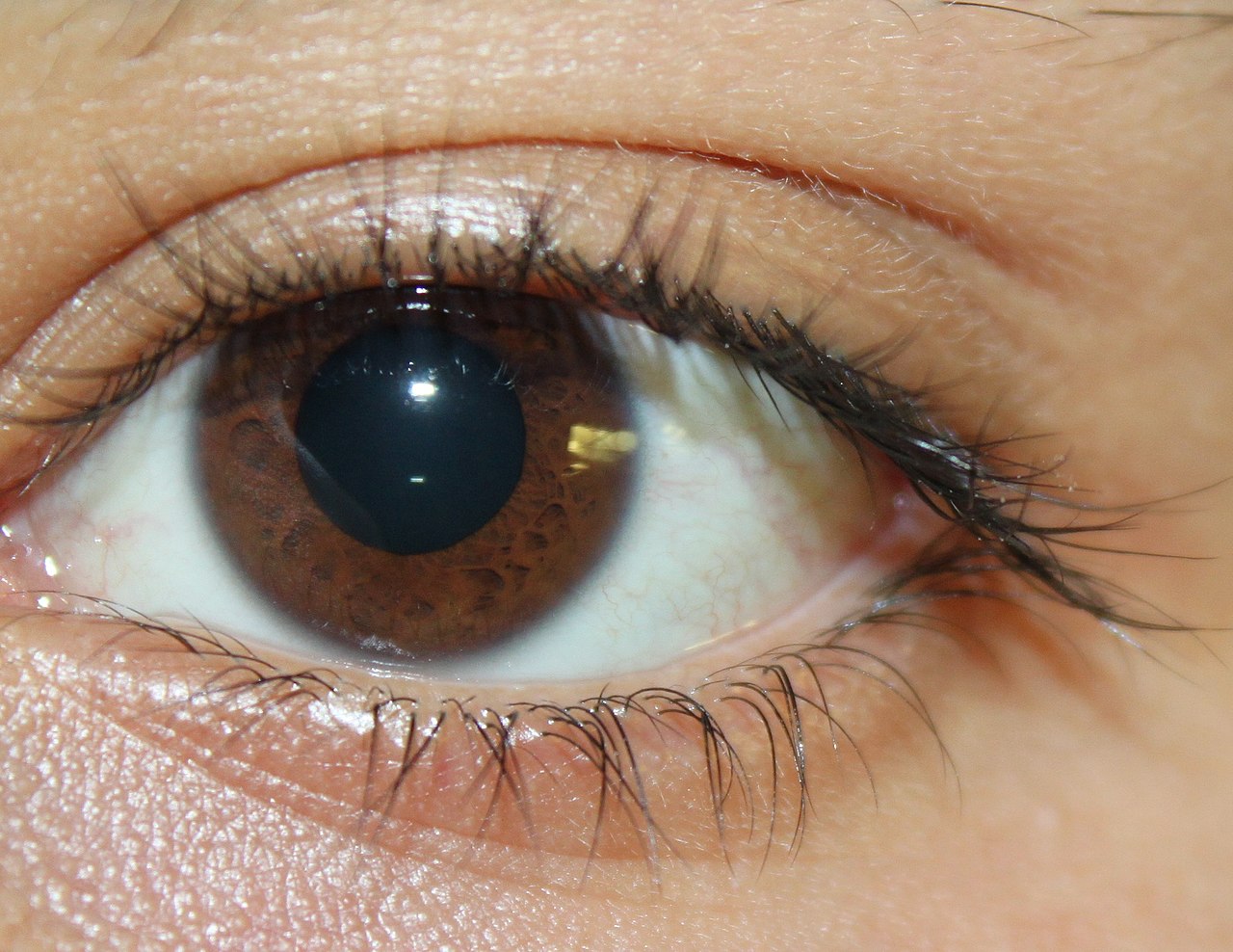





Anatomy and components
The iris consists of multiple layers including an anterior border layer and a fibrous stroma. Two muscle groups lie within it: the sphincter pupillae, which constricts the pupil, and the dilator pupillae, which widens it. A pigmented posterior epithelium and blood vessels supply the tissue; the amount and distribution of pigment contribute to its visible color.
Physiology and control
Pupil size changes in response to light intensity, accommodation (focusing on near objects), and emotional or autonomic factors. Constriction is mediated by parasympathetic pathways while dilation is driven by sympathetic input. By altering aperture size the iris affects retinal illumination, depth of field and visual acuity, and helps protect the retina from excessive light exposure to the retina.
Color and inheritance
Eye color arises mainly from the amount and distribution of melanin in the iris stroma and epithelium. Common human eye colors include brown, blue, green, hazel and gray; rare appearances such as violet or pink are typically associated with lighting, albinism or medical conditions. Color is polygenic and can change slightly with age; variations include sectoral or complete heterochromia.
Clinical significance and uses
Iris disorders include inflammatory conditions such as iritis (anterior uveitis), congenital defects like aniridia or coloboma, and pigment dispersion syndromes. Clinically the iris is examined for signs of disease and trauma. In medicine and technology the iris is notable for biometric identification because its complex pattern is highly individual and stable over time.
History and notable facts
The term "iris" is derived from the Greek word for rainbow and the name of the messenger goddess in classical myth, reflecting the organ's variety of colors. Some alternative practices, like iridology, claim to diagnose systemic illness from iris patterns; these claims lack scientific support. For further overview of eye anatomy see eye references or specialized resources such as eye color studies and technical guides on pupil reflexes at retinal and visual resources.
For authoritative medical guidance consult professional literature; the iris remains a compact, multifunctional structure central to both vision and certain diagnostic or technological applications.
Related articles
Author
AlegsaOnline.com Iris (eye) — structure, function, color, and clinical relevance Leandro Alegsa
URL: https://en.alegsaonline.com/art/48157